§ 2. Main components of the eukaryotic cell
Eukaryotic cells (Fig. 8 and 9) are organized significantly more closely than prokaryotes. Even different stench and for their dimensions (from a number of micrometers to a number of centimeters), and shape, and structural features (Fig. 10).
Rice. 8. Budova cells of eukaryotes. Uzagalne scheme
Rice. 9. Budova's cell for the data of electron microscopy
Rice. 10. Different eukaryotic cells: 1 – epithelial; 2 - blood (e - erythrocytitis, / - leucoitis); 3 - cartilage; 4 - brushes; 5 – smooth m'yazova; 6 - good fabrics; 7 - nerve cells; 8 - cross-smogaste m'azova fiber
The proteo logical organization and the presence of the main components in all eukaryotic cells are the same (Fig. 11).
Rice. 11. Eucaryotic clitina (scheme)
Plasmalemma (the outer clitin membrane). The basis of plasma membranes, as well as other membranes in clitins (for example, mitochondria, plastids, too), is formed by a ball of lipids, which can contain two rows of molecules (Fig. 12). The shards of the lipid molecule are polar (one pole of the stench is hydrophilic, i.e., attracted by water, the other is hydrophobic, i.e., it is exposed to water), those stinks are rotting in order. The hydrophilic kints of molecules of one sphere are directed towards the bic of the aquatic medium - towards the cytoplasm of the clitin, and the other ball - the name of the clitin - in the bik of the interclitinous speech (in the richoclitins) and the aquatic medium (in the uniclitins).
Rice. 12. Budov clitin membranes varying to a common-mosaic model. Proteins of glycoproteins are annealed in the lower ball of lipid molecules, which are called hydrophobic (circles) by their hydrophilic kintsy, and membrane coal is called hydrophobic (wagging lines)
Molecules of proteins are mosaically incorporated into the bimolecular sphere of lipids. From the outer side of the animal clitin to lipids and molecules of proteins in plasma, molecules of polysaccharides are added, which regulate glycolipid and glycoproteins.
Tsya sukupnіst form a ball glycocalyx. Tied to him receptor function plasmalemia (div. lower); in the same way, different speeches can be accumulated in a new one, which victorious clitina. In addition, glycocolix enhances the mechanical stability of plasma membranes.
In clitins, roslin and mushrooms have a clitin wall, which plays a supporting role. In roslin, it is made up of cellulose, and in mushrooms, it is made up of chitin.
The outer clitin membrane performs a number of functions, among them:
♦ mechanical(support, shaper);
♦ barrier-transport(a selection of the penetration of different speeches: the need for clitin of the necessary and the introduction of the inappropriate and the shkidly);
♦ receptor(Designation of various chemical speeches, which have sprung up in uninterrupted proximity to cells; receiving signals from the sight of hormones; recognition of a “foreign” protein by cells of the immune system, etc.).
The exchange of speeches between the clitina and the navkolyshnim middle ground is carried out in different ways - passive and active.
Molecules of water and various ions passively (due to diffusion, osmosis), without consuming clitin energy, come through special pores - tse passive transport. Macromolecules, such as proteins, polysaccharides, inspire the cells of cells, go along the way phagocytosisі pinocytosis from vitratoyu energy - active transport.
The path of phagocytosis is covered by whole cells or large particles (for example, guess about eating in amoebas or phagocytosis by blood cells of bacteria). In case of pinocytosis, there is an exfoliation of fine particles or flecks of rare speech. The most important for both processes are those that slurry of the speech ooze out with an outer membrane, which infuse into the established vacuoles, and then move into the cell cytoplasm glob.
Exocytosis is a process (being also an active transport), directly related to phagocytosis and pinocytosis (Fig. 13). With this help, uncorrupted surpluses of zhі in the simplest, or absorbed in the secretory clitina of biologically active speech, can be removed.
cytoplasm. The cytoplasm is the center of the cells, the surroundings of the plasma membrane, the cream of the nucleus. At її warehouse see main speech (hyaloplasm), organoidsі included.
Hyaloplasm- vyazka homeland, zdatna perebuvati in the camp sol(rarely), or gel(studio-like).
If necessary, the cytoplasm can be reversed from one station to another. For example, in amoeboid Russia (guess the section "Nayprostishi" from the course of zoology), in the course of the adoption of the false foot, there are rapid transitions of the cytoplasm from the gel to the sol and navpaki. The reason for the apparent presence in the cytoplasm is a large number of thread-like molecules from protein actin. If the stench, joining one by one, establishes a trivi- merous line, the cytoplasm is rebuked at the gel station, and if the line falls apart, it can be sol.
In hyaloplasm, there are different kinds of speech - enzymes, proteins, carbohydrates, fats and other, organic and mineral. Here, various chemical processes are involved - the splitting of speech, their synthesis and modifications (changes).
Organoids. These are the permanent components of the clitin with the singing budovoyu and functions, which are carried out in the cytoplasm. Let's talk about organoids of a secret recognition, power to any type of clitin usix eukaryotes. Іz them po'yazane zhittєdіyalnosti remaining. Organoids of special purpose they are less common in clitins of the singing (high-specialized) type - for example, myofibrils in m'yazovyh clitins.
The organoids of a sacrilegious recognition may still be independent of the stench of some kind of organisms of such organisms. Ale among them are seen groups with a membrane (endoplasmic mesh, Golgi apparatus, mitochondria, plastids, lysosomes, vacuoles), and also non-membrane ( ribosomes, clinical center) everyday.
Endoplasmic mesh (EPS). The EPS is composed of membranes and a folded system of tubules and cisterns that permeates the entire cytoplasm of the cell (Fig. 14). Distinguish two types of EPS - shortsі smooth. Ribosomes are attached to the short membranes (from the side of the cytoplasm), but not to the smooth ones.
Rice. 14. Endoplasmic mesh
The endoplasmic line vicons in the eukaryotic cells of the least important functions:
♦ what is the boundary(Submitted the internal obligation of the clergy in the various reactionary spaces);
♦ participation in the synthesis of organic speeches(on the membranes of the short EPS, ribosomes are scattered, and in the smooth - enzyme complexes, which ensure the synthesis of lipids, carbohydrates, etc.);
♦ fate in molding elements of the Golgi apparatus, lysosomes;
♦ speech transport.
Golgi apparatus. Golgi apparatus (AG) - system cisterns(flat vacuoles) bulbashok(Vesicles), spread in close proximity to the nucleus of the clitinum, as if they were settled behind the EPS as a result of the removal of small її fragments (Fig. 15). In case of inflammation of these fragments, new cisterns are blamed on the Golgi apparatus, in case of EPS, different speech tracts are transported, so they take part in the folded folded organic layers (proteins + carbohydrates, proteins + lipids thinly), which should be taken for help of AH between the cells. Biologically active speech either emerges from the cells (for the help of secretory vacuoles through exocytosis), or enters the warehouse of lysosomes (div. lower), which is utvoryuyuyutsya behind the AH.
Rice. 15. Golgi apparatus:
The Golgi apparatus has the following functions:
♦ synthesis biologically active speeches that are vibrated by the clitina;
♦ secretion (vision from cells) of various speeches(hormones, enzymes, speeches, for which there will be a clitin wall, thinly);
♦ the fate of the enlightenment of lysosomes.
Mitochondria. Mitochondria are present in all types of eukaryotic cells (Fig. 16). The stench may look either rounded bodies, or sticks, or rather - threads. The diameters are kolivayutsya in 1 to 7 microns. The number of mitochondria in klitz should be from a few hundreds to tens of thousands (in the great simplest).
Rice. 16. Mitochondria. In the mountains - mitochondria (a) near the sciatic canals, visible under light microscopy. Below - a trivimiral model of the organization of mitochondria: 1 - Christi; 2 - outer membrane; 3 - inner membrane; 4 - matrix
Mitochondrion is covered with two membranes - old-fashionedі internal, mizh yakimi rozashovano intermembrane space. The inner membrane satisfies the impersonal vp'yachuvan - Christ, either plates or tubes. Such an organization ensures the great area of the inner membrane. Enzymes grow on it, which provide the transformation of energy, which is stored in organic speeches (in carbohydrates, lipids), into the energy of ATP, which is necessary for the life of the cells. Also, the function of mitochondria is the fate of energy clinical processes. The very large number of mitochondria is attached to it, for example, to the m'azovim clitins, like a great robot.
Plastidi. In growing plants, there are special organoids - plastids, which often have a spindle-like or rounded shape, sometimes folded. Three types of plastids are distinguished - chloroplasts (Fig. 17), chromoplasts and leukoplasts.
Chloroplasty they are ablaze with a green color, a kind of pigmentation. chlorophyll, what will secure the process photosynthesis, to the synthesis of organic speech from water (H 2 O) and carbon dioxide (CO 2) with the energy of sony light. Chloroplasts are more important in leaf clitins (in higher growths). The stench is formed by two parallel membranes, one by one, which are separated from chloroplasts. stroma. The inner membrane satisfies the numerical flattening of the little balls. thylakoids, yakі folded in piles (for a stack of coins) - edges - and lie at the stroma. Itself in thylakoids and chlorophyll.
Chromoplasts they signify yellow, orange and red colors of rich flowers and fruits, which are present in the houses of a great number. The main pigments at the warehouse carotene. Functionally, the recognition of chromoplasts is more pronounced in the color of the irradiated creatures, which ensure the sawing of the tickets and the width of the bottom.
Rice. Fig. 17. Plastidi: a – chloroplasts in elodea leaves, visible under light microscopy; b - scheme of the inner chloroplast with facets, which are hundreds of flat sacs, splayed perpendicular to the surface of the chloroplast; c - a larger report scheme, which shows the anastomotic tubules that work around the fan chambers
Leukoplasty- all bezbarvnі plastidi, scho to be near the clitins of the underground parts of the roslins (for example, near the bulbs of potatoes), only that core of the stem. In leukoplasts, the main rank is the absorption of glucose and starch and the accumulation of yoga in the organs of the roslin, which is stored.
Plastids of one kind can transform into another. For example, during the autumn change in the color of the leaves, chloroplasts are transformed into chromoplasts.
Lysosomes. Qi organoids may look like bulbs, sharpened with a membrane, up to 2 microns in diameter. The stink of a sprat of dozens of enzymes that break down proteins, nucleic acids, polysaccharides and lipids. The function of lysosomes is the fate in the processes of internal cleavage of folded organic sprouts (for example, pharyngeal rhizomes or rhechovins, which "repaired" clitinic components). Lysosomes engulf with phagocytic (or pinocytic) vacuoles, making herbal vacuoles.
Adopted by lysosomes, it is released from the kidneys in the cisterns of the Golgi apparatus.
Ribosomes. Ribosomes (Fig. 18) are present in clitins, both eukaryotes and prokaryotes, and shards play an important function in biosynthesis of proteins(div. division 5). In skin cells there are tens, hundreds of thousands (up to a few million) of these other rounded organoids.
Rice. 18. Scheme of a ribosome that sat on the membrane of the endoplasmic membrane: 1 – small subunit; 2 - tRNA; 3 – aminoacyl-tRNA; 4 - amino acid; 5 - large subunit; 6 - membrane of the endoplasmic membrane; 7 – synthesis of polypeptide lanciug
The ribosome is made up of two nerve subunits (parts). The stench settles down and merges, “hoaring” the informational RNA, during the process of protein molecule synthesis. The warehouse of ribosomes includes various proteins and ribosomal RNA.
Clitinni included. This is the name of non-permanent components in clitiny, which are present in the main speech of cytoplasm like grains, granules and speckles. The inclusions can be sharpened by a membrane, or they will not be irritated by it.
Functional wear has three types of inclusion: spare living speeches(starch, glycogen, fats, proteins), secretory inclusion(speech, characteristic of salivary clitin, which are produced by them, - hormones of saliva internal secretion toshcho) inclusion of special recognition(In highly specialized cells, for example, hemoglobin in erythrocytes).
§ 3. Organization of the clitin nucleus. Chromosomes
The clitin nucleus (div. fig. 8 and 9) is more important in the life of the clitin, but it can serve as a treasure trove of decay information that is stored in the chromosomes (div. below).
The core is surrounded by a nuclear shell, which makes it stronger (karyoplasm) type of cytoplasm. Shell and two membranes separated by an interlayer. Offensive stinks are permeated with numerous pores, zavdyaki like a possible exchange of speech between the nucleus and cytoplasm. In the nucleus of the clitin, in most eukaryotes, there are types 1 to 7 nuclear They involve processes for the synthesis of RNA and tRNA.
Main components of the kernel chromosomes, digestion of DNA molecules and other proteins. At the light microscopy, the stench of the good remembers only the next hour of the client's pod (Mitosis, meiosis). In cells that do not divide, the chromosomes look like long thin threads, spread over the entire volume of the nucleus.
Under an hour, the chromosomal strands settle down the thin spirals, due to which they become visible (behind the help of a powerful microscope) in the form of sticks, “hairpins”. All of the genetic information is distributed between the chromosomes of the nucleus. The process of breeding was revealed the following regularities:
♦ in the nuclei of somatic clitins (that is, the clitin of the body, non-state) in all individuals of the same species, the same number of chromosomes is located, which set of chromosomes(Fig. 19);
Rice. 19. Chromosomes of different species of roslin and creatures, depicted in one scale: 1,2 - amoeba; 3,4 - diatom algae; 5–8, 18.19 – green algae; 9 - fly agaric; 10 - linden; 11–12 – Drosophila; 13 - salmon; 14 - skerda (homeland of folding flowers); 15 - roslina from the aroid family; 16 - panicle-chubatka; 17 - comacha from the locust family; 20 - water bug; 21 - flower bug; 22 - amphibious ambistoma; 23 - scarlet (family of lilies)
♦ for the dermal species, a characteristic number of chromosomes is characteristic for each number (for example, humans have 46 chromosomes, Drosophila fly - 8, roundworm - 4, river cancer - 196, horse - 66, corn - 104);
♦ chromosomes in the nuclei of somatic cells can be grouped in pairs, which are named homologous chromosomes on the basis of their imitations (for household and functions);
♦ in the nuclei of state clitins (gametes) from the skin pair of homologous chromosomes, there is less than one, so the overall set of chromosomes in two-witches is smaller, lower in somatic clitins;
♦ a single set of chromosomes in state cells is called haploid day i is denoted by the letter n, and somatic - diploid day(2n).
It is clear from the foregoing that the skin pair of homologous chromosomes is assigned to the union of the father's and maternal chromosomes during the fusion, i.e., the fusion of the state cells (gametes). First of all, with the established clitins from the skin pair of homologous chromosomes, only one gamete is consumed.
Chromosomes rіznih homologous pairs are dependent on sizes and shape (Fig. 20 and 21).
Rice. 20. Budova and type of chromosomes: a – old appearance 1 – cetromir; 2 - short shoulder; 3 - dove shoulder); the internal structure of the same chromosome (1 - centromir; DNA molecules); c - type of chromosomes (1 - single arm; different arm; 3 - equal arm: X - arm, V - centrifuge)
Rice. 21. The chromosome is made up of DNA and proteins. The DNA molecule replicates. Two identical subfolding strands of DNA are occluded in the region of the entromer. Qi copies are recreated on the same chromosomes later, every hour under the cells
In thіlі chromosomes are seen the first constriction (called the center), until the threads are attached spindle under. Divide a chromosome into two shoulder. Chromosomes can be equal-armed, different-armed and one-armed.
Section 5. Exchange of speeches
§ 1. Metabolism as a unity of asimilation and dissimilation
All cells and living organisms are viable systems, so that the stench peresbuvayut at the station of the constant exchange of energy and speeches from the most important medium. Є vіdkritі system y in inanimate nature, that їх іх іхування yakіsno vіrіznyаієєі in living organisms. Let's look at such a butt: a piece of native sirka, which burns, is known in the exchange camp with the most important medium. At the yogo mountain, O 2 is clayed, and SO 2 is seen that energy (it looks like heat). However, at the same time, the body collapses like a physical body, losing its original structure.
For living organisms, the exchange with a superfluous medium is manifested by mental conservation, support of their structural organization by the self-renewal of all speeches and components, from which stinks are added.
The exchange of speech (metabolism) is the sequence of processes that take place in living organisms (regeneration, transformation, accumulation and vision of speech and energy), which ensure their life, development, growth, creation. In the process of exchange of speech, splitting and synthesis of molecules occurs, which enters the warehouse of clitin; renewal of clitinic structures and intercellular speech.
Metabolism is based on mutual processes assimilation(anabolism) that disimilation(Catabolism). With asimilation (plastic exchange), the synthesis of folding speeches from simple ones is observed. The very beginnings of this are created by all organic speech in clitin, which is necessary for inducing structural components, enzyme systems, too. Asimilation is always building up from vitratoy energy.
In the course of dissimilation (energy exchange), folded organic speeches are split into simple inorganic ones. When you see the energy, as if stained by the clitinum on the viconnance of the various processes, which will ensure life (the synthesis and transport of speeches, the mechanical work is just fine).
All living organisms can be divided into two groups: autotrophyі heterotrophy, yakі vіdrіznyayutsya dzherel energy and nebhіdnyh rechovina for the security of their lives.
Autotrophy- organisms that synthesize from inorganic speeches organically with vicarious energy of sony light (like phototrophy- growths, cyanobacteria or energy, obtained during the oxidation of mineral (inorganic) speeches (such as chemotrophy– serobacteria, salizobacteria and others). Otzhe, the stench of the building independently create the necessary speeches for your life.
§ 2. Dissimilation in anaerobic and aerobic organisms
Organisms can be divided into two groups according to the nature of dissimilation - aerobiі anaerobic. Aerobi (type Greek. bіs- repeat) require a free sour for life. In anaerobes (Greek. ats- Negative part) there is no need for it. Their dissimilation develops along a path of wandering - acid-free, enzymatic splitting of organic speech with more simple organic speeches and visions of energy. For example:
♦ lactic fermentation:
C 6 H 12 O 6 + 2H 3 PO 4 + 2ADP → 2F H + 2ATP + 2H 2 O;
♦ alcohol fermentation:
C 6 H 12 O 6 + 2F H + 2ADP → 2C 2 H 5 OH + 2CO 2 + 2ATP + 2H 2 O.
Speech, which are settled during wanderings, is organic and, then, revenge is still rich in energy.
Rice. 22. Mutual assimilation and dissimilation in autotrophic and heterotrophic organisms
In aerobic organisms, in the process of breathing in mitochondria, there is an increase in the splitting of organic speech (with vicarious Pro 2) to the most energy-efficient end products of CO 2 and H 2 Pro and significantly more energy is seen:
H 6 H 12 0 6 (glucose) + 0 2 > 6C0 2 + 6H 2 0 + energy (38 ATP molecules are synthesized per day).
Let's take a look at the detailed schemes of metabolism in autotrophic and heterotrophic aerobic organisms (Fig. 22).
Asimilation. The most important processes - photosynthesisі biosynthesis of proteins.
Chapter 1
FOUNDATIONS OF CLIENT PHYSIOLOGY
I. Dudel
plasma membrane . Creatures of cells are surrounded by a plasma membrane (Fig. 1.1). On the її budovі, even similar to the richness of the inner clitin membranes, we can hardly hear the report. The main matrix of the membrane is composed of lipidiv, head rank phosphatidylcholine. Lipids are formed from the head hydrophilic group, up to the adjunct of two hydrophobic in carbohydrate lanciugs. In water, lipids spontaneously form a two-ball plume with a thickness of 4-5 nm, in some hydrophilic groups, they grow to a watery middle, and hydrophobic, in carbohydrate, lances, they grow in two rows, satisfying an anhydrous lipid phase. Cell membranes with lipid bilayers of this type can also be used for glycolipids, cholesterol and phospholipids (Fig. 1.2). The hydrophilic part of glycolipids is filled with oligosaccharides. Glycolipids are regularly distributed on the outer surface of the plasma membrane, and the oligosaccharide part of the molecule is oriented similarly to a hair embedded in the middle of the middle. The expansion of the middle phospholipids in myzhe equal number of cholesterol molecules stabilize the membrane. Rozdil different lipids in the inner and outer balls of the membrane is not the same, and wind at the borders of one ball є plots, which concentrate around the same lipids. Such a nervous rozpodіl,
Rice. 1.1. Schematic baby clitiny, which depicts the most important organelles
maybe, maybe, it’s still unclear, functionally.
The head functional elements, zanurenny in the porous inert lipid matrix of the membrane, є proteins(Fig. 1.2). Protein by weight becomes 25 to 75% in different membranes, but the number of protein molecules is richer, lower lipid, 50% by weight is equivalent to the ratio: 1 protein molecule per 50 lipid molecules. Some proteins penetrate the membrane from the outside to the inner surface, others are fixed in one ball. Protein molecules sound oriented so that their hydrophobic groups are numbed at the lipid membrane, and the polar hydrophilic groups on the surface of the membrane are numbed at the aqueous phase. Rich in proteins of the outer surface of the membrane - glycoproteins; їх hydrophilic saccharide groups of animals in the subacute middle.
Membrane systems of internal cellular organelles .
Approximately half of the cell volume is occupied by organelles isolated from the cytosol by membranes. The upper surface of the membranes of the intracellular organelles is taken 10 times over the surface of the plasma membrane. The most expanded membrane system - endoplasmic reticulum, is a measure

Rice. 1.2.Schematic representation of the plasma membrane. The proteins are anchored at the phospholipid bispheres, moreover, the deacons from them penetrate the bispheres, although others are only anchored on the outer or inner ball.
strongly coiled tubules or bell-shaped curly structures; large spaces of the endoplasmic reticulum with ribosomes; such a reticulum, having taken off the name of the granular, or short (Fig. 1.1). Golgi apparatus it is also formed from membrane-bound lamellae, in which bulbs or vesicles are introduced (Fig. 1.1). Lysosomes and peroxisomes– only small specialized vesicles. In all these various organelles, the membrane and space, which are surrounded by it, contain specific sets of enzymes; in the middle of the organelles, special exchange products are accumulated, victorious implementation of various functions of the organelles.
Coreі mitochondria note that the skin of these organelles is sharpened with two membranes. The core is viable for the kinetic control of metabolism; folding of the inner membrane of mitochondria - the site of oxide metabolism; here, for the oxidation of pyruvate or fatty acids, a high-energy half of adenosine triphosphate (ATP, or ATP) is synthesized.
cytoskeleton . The cytoplasm, which is the source of organelles, cannot even be considered amorphous; її penetrating the mesh of the cytoskeleton. The cytoskeleton is composed of microtubules, actin filaments and intermediate filaments (Fig. 1.1). microtubules may have an actual diameter of about 25 nm; the stench settles down like a great polymer, after the selection of tubulin protein molecules. Actin filaments fast-moving fibers that grow in the membrane ball and in all cells - it is important to take part in the processes that are tied with ruh. Intermediate filaments stacked from blocks of different chemical storage in different types of cells; stinks make different connections between the two other elements of the cytoskeleton. The organelle and the plasma membrane are also connected with the cytoskeleton, which not only ensures the maintenance of the shape of the cell and the position of the organelle in them, but also the change in the shape of the cell and its fragility.
Cytosol . Nearly half of the clitinum is occupied by cytosol. About 20% (per weight) of the wines is added up for the protein, the price is higher for the gel, the lower water price. Small molecules, including organic and inorganic ioni, differences in the water phase. Mіzh kіtinoyu and navkolishnіm sredovischem (zaklіtinny expanse) vіdbuvaetsya exchange of ions; cі exchange processes will be looked at at the offensive division. The concentration of ions in the post-acute space is measured with a significant accuracy to the constant equal; the intracellular concentration of skin ion ions may also be specific, which may vary in such a posture (Table 1.1). The largest cation in the postacute medium is Na+ in clitiny yogo, the concentration was higher lower, 10 times lower. Navpaki, in the middle of the clitinum the highest concentration of K + position of the clitinum is lower by an order of magnitude. The largest gradient between post-clitin and intra-clitin concentrations is observed for Ca 2+ concentration of free ions in the middle of the clitin at 10,000 times lower, lower after it. Not all of them are distributed in the cytosol, their part is adsorbed on proteins or deposited in organelles. For example, in the case of Ca 2+, they are rich in numbers, lower in number. The majority of proteins in the cytosol are enzymes, with the participation of which they take part in the immaterial processes of intermediate metabolism: glycolysis and gluconeogenesis, synthesis of a number of amino acids, protein synthesis on ribosomes (Fig. 1.1). In the cytosol, there are also droplets of lipids and granules of glycogen, which serve as reserves of important molecules.
Table 1.1.Internal post-clitin concentrations of ions in malignant clitins of homoiothermic creatures. BUT – - “high molecular weight clitin anions”
|
intracellular concentration |
Pozaklіtinna concentration |
||
|
Na+ |
12 mmol/l |
Na+ |
145 mmol/l |
|
155 mmol/l |
Up to + |
4 mmol/l |
|
|
Ca 2+ |
10 -7 10 -8 mmol/l |
Ca 2+ |
2 mmol/l |
|
Z l - |
4 mmol/l |
Z 1 - |
120 mmol/l |
|
HCO 3 - |
8 mmol/l |
HCO 3 - |
27 mmol/l |
|
A- |
155 mmol/l |
Other cations |
5 mmol/l |
|
Quiet potential -90 mV |
|||
1.2. Exchange of speeches between clitin and navkolishnim middle
We briefly described the budovu clitiny, so that we could describe the vicariate to understand the basics of clitinous physiology. The clitina at any time cannot be taken into account by static denunciations, but here there is a constant exchange of speech between different internal clitin compartments, as well as between compartments and the present middle. The structures of the clitin are in a dynamic balance, and interchange of the clitin between itself and the outer core, which is necessary for the mind to support the life of a functioning organism. We have seen the fundamental mechanisms of such an exchange. In advancing divisions, the mechanisms will be examined at the appendage to the nerve cell and its functions;
however, these mechanisms themselves underlie the functioning of all other organs.
Diffusion.The simplest process of moving speech is diffusion. In different regions (both gases), atoms and molecules move freely, and the difference in concentrations is leveled off by diffusion. Let's look at two obsyags, filled with natural gas (Fig. 1.3), in which speech concentrations can be s 1 i s 2 that is divided by a sphere with a surface area, and that by a torso d. Speech flow m per hour t be described the first law of diffusion Fik:
dm/ dt= DA/ d ( C 1 -З 2) =DA/ dD C(1)
de D-coefficient of diffusion, constant for this speech, rozchinnik and temperature. Have a bigger look for retail concentrations dc on dx
dm/dt= -D A dc/dx,(2)
flow through the passage A proportional to the concentration gradient dc/dx . The “minus” sign blames y equal to the fact that the change in concentration y is directly x-negative.
Diffusion is the most important process, because most of the molecules in water areas move to small distances. It is necessary to be carried and up to the last moment in the clitiny leaves, the fragments of diffusion do not permeate the membranes. Anonymous speech can freely diffuse through lipid membranes, especially water and gases, such as 2 and 2. Zhirorozchinnі

Rice. 1.3.Kilkisna scheme of diffusion. Two open spaces separated by a balloondand square BUT. C; - high concentration of particles in the left part of the oblique, C: - low concentration of particles in the right particles, erysipelas-Concentration gradient in diffusion sphere. Diffusion potik dm/dt-div. equalization (1)
speech also diffuses well through the membranes; it is necessary to push and polar molecules to reach a small size, such as ethanol and sechovin, just like zukri pass through the lipid ball by force. The density of lipid spheres is practically impenetrable for charged molecules, including those for inorganic ions. For non-electrolytes, diffusion equalization (1) should be changed, bringing the characteristics of the membrane and diffuse speech into one parameter-penetration (P):
dm/dt=P AD with.(3)
On fig. 1.4 look up permeability (P) of the lipid membrane for different molecules
Diffusion through membrane pores . The plasma membrane (and other cellular membranes) is not only penetrating for speech, which diffuses through the lipid sphere, but also for rich ions, sucrose, amino acids and nucleotides. Qi of the speech dolayat the membrane through the pores, transport proteins, screw up the membrane. The middle of such proteins is a channel filled with water with a diameter of less than 1 nm, through which small molecules can diffuse. The stench collapses along the concentration gradient, and even though the stench carries a charge, its ruh channels are also regulated by the membrane potential. Membrane channels may be vibrating.

Rice. 1.4.Penetration of piece lipid beads for various speeches
According to the type of molecules, which can pass through them, for example, potassium, sodium and calcium channels, skin from some non-penetrating practical for any ion, specific cream. Taka vibrancy bound by a charge or structure, the bonding site at the walls of the channel, which facilitates the transport of a specific molecule and avoids the penetration of other tracts through the channel (small. 1.5, A) .
For behavior membrane ion channels easily posterigati, oskіlki strum, scho vinikaє in rusіonіv, you can vymіryati, moreover, navіt for a single channel. It is shown that the channels spontaneously and with a high frequency change their state from open to closed. The potassium channel is characterized by strum pulses with an amplitude of close to 2 pA (2 10 -12 A) and a trivality of a few milliseconds (div. Fig. 2.12, page 37) [Z]. During this period, tens of thousands of ions pass through it. The transition of proteins from one conformation to another is carried out by X-ray diffraction, Mössbauer spectroscopy and nuclear magnetic resonance (NMR). Proteins, in this order, are already dynamic, crumbling structures, and the channel that passes through the protein is not just a thick, water-filled tube (small 1.5, A), but of molecular groups and charges, which are easily collapsing. Qia dynamic characteristic of the channel is displayed in energy profile channel, shown in fig. 1.5, B. Here, along the abscissa axis, the channel is represented from the outside range with an ion concentration of 0 and a potential of 0 to the internal range with a concentration of 1 and a potential E. On the y-axis

Rice. 1.5.A. Scheme of the protein, which regulates the potassium channel, numbing the lipid bilayer of the plasma membrane. Chotiri negative charge fixed on the "stіntsi" channel. B. Schematic energy profile of the channel shown in fig. A. On the y-axis, you can see the value of the kinetic energy required to pass through the channel; along the abscissa axis - stand between the inner and outer surfaces of the membrane. The energy minimums induce the channels to positively charge the ions and fix the negative charges at the channel wall. Energetic maxima indicate a shift in diffusion in the channel. It is believed that the conformation of the channel protein spontaneously oscillates; options for energy profiles depicted with strong and dashed lines; tsі oscilyatsії znachnym іroy it is easier to link the ions with the help of the energy bar'єru (behind the changes)
representation of the equal energy of the ion in the communication areas of the channel; The peak on the graph represents the barrier of penetration, which energy is responsible for overturning for penetration through the channel, and the “failure” of the graph is a similarly stable station (linking). Regardless of the transition at the energy peak, the wine can penetrate through the channel, as the energy profile spontaneously changes cyclically; ion, in such a manner, can quickly appear "on that bek" of the energy peak and can continue to move at the client. Fallow in the charge, rozmiriv that degree of hydration of the ion and yogo zdatnosti zv'yazuvatisya z_ structures of the walls of the channel energy profile of the channel varies for different ions, which can explain the vibrancy of other types of channels.
Diffusion rіvnovaga ionіv . Diffusion of various ions through the membrane channels is small to lead to the absorption of water in the concentrations between the interstitial media. Yak, prote, can be seen from the table. 1.1, such discretion is conserved, then, may be jealous between diffusion and other processes of transport through the membrane. In different ions, the diffusion charge is injected into the diffusion line. Diffusion of uncharged molecules is ensured by concentration differences dc , and if the concentrations rise, the transport will be wetted. On charging the particles, an electric field is injected. For example, if the potassium ion comes out of the cell following its concentration gradient, it carries one positive charge. In this way, the inner clitinum becomes more negatively charged, after which the difference in potentials is caused on the membrane. The internal negative charge transfers new ions to potassium from the cells, and these ions still appear from the cells, increasing the charge on the membrane even more. Potik ion in potassium is attached, if the electric field compensates for the diffusion pressure due to differences in concentrations. They continue to pass through the membrane, but at equal distances in both directions. Also, for a given difference in the concentration of ions on the membrane equal potential E ion when the flow of ions through the membrane is attached. An equal potential can be easily assigned for help Rivnyannia Nernst:
Eion= RT/ zF* lnOut/ C in(4)
de R -gas postina, T-absolute temperature, z - ion valence (negative for anions) Out - postacute concentration of the ion, C in - Intracellular ion concentration, F Faraday number. If we represent in equal constants, then for body temperature (T = 310 K) equal potential for potassium ions E K dorivnyuє:
Ek= –61 mB log /(5)
Yakscho [ K + out ]/[ K + in ] = 39, as shown in the table. 1.1, also
Ek = -61 m B log 39 = -97 mV.
It’s true, it’s been revealed that all clitins can membrane potential; in m'yazovyh clitinah savtsiv rіven become close to -90 mV. In fallow waters and water concentrations of ions in cells, the membrane potential at the boundaries of waters can be -40 to -120 mV. For clitiny at the pointed eye butt (Table 1.1) peace potential, a ratio of about -90 mV indicates that potassium ion fluxes through membrane channels are approximately equal. It’s not surprising, the shards in the membrane, which lie, the opening of the potassium channels is the most comfortable, tobto. the membrane is the most permeable for potassium ions. The membrane potential, however, is determined by the flows of those other ions.
The lightness, with which uncharged particles can diffuse through the membrane, is predominantly presented in equal (3). Penetration for charged particles Descho folded equals:
P= m RT/ dF(6)
de m- Roughness of the ion in the membrane, d - The thickness of the membrane, a R , T and F - in the thermodynamic post. As such, the values of penetration for various ions can be changed for the development of the membrane potential Em , if the ions of potassium, sodium and chlorine pass through the membrane at the same time (with penetration R K, P Na and P Cl obviously). When it is transferred, the potential drops in the membrane equally, so the field strength is constant. Whose mind gets stuck Goldman's leveling, or the standing field's leveling :
Em= R T/F * ln(P K + P Na + P Cl )/ (P K + P Na + P Cl )(7)
For larger clitin membranes P K about 30 times higher, lower R Na (Div. also section 1.3). Vіdnosna value PCl highly variable; for rich membranes PCl small in povnyannі s R K however, for others (for example, in skeletal m'yazakh) PCl significantly higher, lower R K.
Active transport, sodium pump . In the front section, the passive diffusion of ions and the membrane potential are described, which are satisfied later on, when setting the internal and secondary concentrations of ions. However, in the result of this process, the concentration of ions in the middle cells does not stabilize automatically, membrane shards
potential less more negative, lower E K , i richly - povnyano s E Na (close to +60 mV). Zavdyaki diffusive internal concentrations of ions, taking potassium and sodium, are due to zakletinnymi. The stability of the gradient of ions is reached by additional active transport: membrane proteins carry ions through the membrane of the opposite electric and (or) concentration gradients, spozhivayuschie for this metabolic energy. The most important process for active transport is the purpose of the robot Na/K -pump, which is practical for all customers;
the pump pumps sodium ions out of the cells, simultaneously pumping potassium ions into the middle of the cells. In this way, a low intracellular concentration of sodium ions and high potassium is ensured (Table 1.1). The concentration gradient of sodium ions on the membrane has specific functions related to the transmission of information in the appearance of electrical impulses (div. Rev. 2.2), as well as to the support of other active transport mechanisms and regulation of the environment (div. 2.2). It is not surprising that more than 1/3 of the energy, which is saved by the cell, is spent on the Na / K-pump, and in some of the most active cells, up to 70% of the energy is spent on the robot.
Na/K-transport protein є ATPase. On the inner surface of the membrane, it splits ATP into ADP and phosphate (Fig. 1.6). The energy of one ATP molecule is generated for the transportation of three sodium ions from cells at the same time as two potassium ions to cells, i.e., in total, one positive charge is generated per cycle from cells. Thus, the Na/K-pump is electrogenic(Creates an electrical stream across the membrane) to increase the electronegativity of the membrane potential by approximately 10 mV. The transport protein is used for high speed operation: from 150 to 600 sodium ions per second. The amino acid sequence of the transport protein is the prototype of the understanding of the mechanism of the folding exchange transport. This process is described for the help of energy profiles transferred by proteins to sodium or potassium ions (Fig. 1.5.5). Behind the nature of the change in profiles, caused by permanent changes in the conformation of the transport protein (the process that leads to energy gain), one can judge the stoichiometry of the exchange: two potassium ions are exchanged for three sodium ions.
Na/K pump, insulated Na+ /K + -deposited membrane ATPase, specifically inhibited by cardiac glycoside ouabain (strophanthin). Shards of the Na / K-pump robot with a rich chemical reaction, won, like all chemical reactions, a significant world to lie in the temperature that

Rice. 1.6.Diagram of the Na/K-ATPase pump (anurated into the lipid bilayer of the plasma membrane), so that in one cycle it can bring three Na + ions from the cell against the gradient of the concentration and bring two K ions from the cell + . In the process, one ATP molecule is split into ADP and phosphate. On the scheme, ATPase is shown as a dimer, which is composed of large (functional) and small subunits; the membrane has it as a tetramer, the solutions of two great and two small subunits
demonstrated on a small 1.7. Here potіk ionіv natіyu z m'azovih clitin indications at the hour; practically equivalent to the flow of sodium ions mediated by the Na/K-pump robot, so that the passive flow of sodium ions is against the concentration gradient and the marginal potential. If the drug is cooled by approximately 18 ° C, then the sodium ion flux from cells will change rapidly in 15 times, and once heated, it will increase to the cob level. Such a change in the flow of sodium ions from cells in a kilka times more, lower those that would lead to a temperature-delayed diffusion process or a simple chemical reaction. A similar effect is expected if the stock of metabolic energy is exhausted through the disruption of dinitrophenol (DNF) (Fig. 1.7.5). Later, the flow of sodium ions from the cell is protected by an energy-storage reaction - an active pump. The second characteristic of the pump is the order of the significant temperature and energy deposits - the presence of the level of the increase (as in all other chemical reactions); This means that the speed of the pump operation cannot be increased indefinitely with increasing concentrations of ions that are transported (Fig. 1.8). At a glance, passively diffuse speech grows in proportion to the difference in concentrations, according to the law of diffusion (equals 1 and 2).
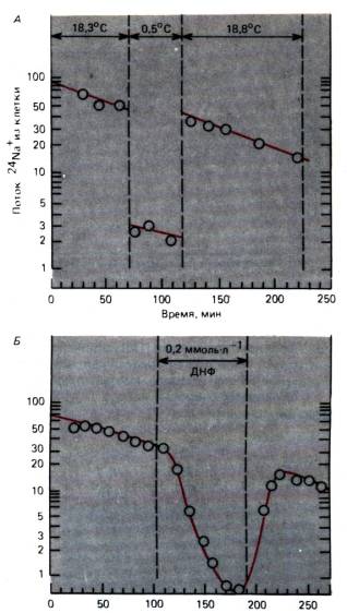
Rice. 1.7. A, B. Active Na transport +. All ordinates: Potik radioactive 24 Na + cells (Imp./HV). Abscissa: hour on the cob experiment. BUT. Klitina was cooled from 18.3 °С to 0.5 °С; sweat Na+ from clitiny during the whole period of galvanization. B. Impairment of the flow of Na + from cells with dinitrophenol (DNF) at a concentration of 0.2 mmol / l (with changes)
Crim Na/K-pump plasma membrane to replace one pump - calcium; tse pump vodkachuє іoni calcium (Са 2+) from clitin and take part in podtremtsі їх vnutrіshnоklіtinnoї kontsentratsії at a very low level (tab. 1.1). Calcium pump is present in an arc of high thickening in the sarcoplasmic reticulum of malignant cells, as they accumulate calcium ions due to splitting of ATP molecules (div. ch. 4).
Na/K-pump injection on membrane potential and cell volume . On fig. 1.9 shows the different components of the membranous strum and induces internal cellular concentrations of ions, yak

Rice. 1.8.Spivvіdnennia between the transport of molecules and their concentration (at the entrance to the channel or in the pump connection) during diffusion through the channel or pumping transport. Remaining at high concentrations is observed (maximum fluidity, Vmax ) the value on the abscissa axis, which shows half of the maximum fluidity of the pump ( Vmax /2), є equally important concentration Before m

Rice. 1.9.Scheme showing Na + concentration , K+ ta Cl- in the middle of the posture of the clitin and the path of penetration of these ions through the clitin membrane (through specific ion channels or with the help of Na / K-pump. With these concentration gradients, equal potentials E Na , E K and E С l – equal, membrane potential Em = – 90 mV
take care of їhnє іsnuvannya. Through the potassium channels, a stream of potassium ions is prevented from leaving, since the membrane potential is more electropositive, lower than the equal potential for potassium ions. The total conductivity of sodium channels is richly lower, lower than potassium, tobto. sodium channels are richer, lower potassium at the potential to calm; However, Kl_tin is approximated by the stilki, Skіlki to enter the zhoshi Іonіv Kalіu, in the same time for Difuzіїv, Natrіu in Klіtin Nechіdnі G. Gradієnti Concentzії likea. The Na/K-pump provides ideal compensation for passive diffusion streams, so that sodium ions are transferred from cells and potassium ions are transferred into them. In this way, the pump is electrogenic for the account of the difference in the number of charges transferred from the cell and from the cell, which, with normal fluidity of the robot, creates a membrane potential of approximately 10 mV is more electronegative, lower than vin utvoryuvavsya only for passive ions flows (div. rіvnyannya 7). As a result, the membrane potential approaches the potassium equal potential, which changes the potassium ion coil. Na activity/K-pump is regulated by the internal concentration of sodium ions. Schwidkіst Roboty Pumps Usilnunyuyuyu at the Znigensian concentrations Іonіv Natroju, Shah, pіdlagut the Vyplennia z Klіtini (Fig. 1.8), so Shah's robot of the pump Tu Potіk Іonіv Natroju in the middle of Klіtini to vrolitnayn the one, pіdtriyuyi internaloklіtinna concentricіyu natroju Іonіv on Rivnі is about 10 mmol / l.
In order to improve the efficiency of inter-pumping and passive membrane strums, it is necessary to have more Na/K-pump molecules, lower channel proteins for potassium and sodium ions. When the channel is open, tens of thousands of ions pass through the new one in a few milliseconds (divine more), and the shards of the channel sound like a few times for a second, more than 10 5 ions pass through the new one in an hour. A single pumping protein moves hundreds of sodium ions per second, hence, the plasma membrane is responsible for approximately 1000 times more pumping molecules than channel molecules. The reversal of channel strums in calm showed the presence of one potassium and one sodium hydroxide channel per 1 µm 2 of the membrane in the middle; Why is it obvious that this space can have close to 1000 molecules of the Na/K-pump, that is. between them to become the average 34 nm; the diameter of the pump protein as a channel becomes 8-10 nm. Thus, the membrane is heavily loaded with pumping molecules.
The fact that the flow of sodium ions into the middle of the cell, and potassium ions from the cell is compensated by the operation of the pump, is another consequence that affects the saving of a stable osmotic vice fast oblige. The middle cells have a high concentration of great anions, the main rank of whites (A - in Table 1.1), which is not able to penetrate through the membrane (or it is more appropriate to penetrate through it) and that is the fixed component of the middle cells. Schob ur_vnovazhiti charge of tsikh anion_v, p_vna k_lk_st kat_onіv. The pumps of the Na/K-pump with cations are mainly potassium ions. A slight increase in the internal clitin concentration of ions could be observed only with an increase in the concentration of anions in the afterflow of C1 - behind the concentration gradient at the clitin (Table 1.1), but the membrane potential against that. Entrance strum Cl- beware of only dots, the docks will not reach an equal potential for chlorine ions; It is worth considering, if the gradient of ions to chlorine is practically opposite to the gradient of ions of potassium, then they are negatively charged to chlorine (equal 4). In this way, a low internal concentration of ions in chlorine is established, which results in a low posterior concentration of ions in potassium. The result is the defecation of the total number of ions in the clitin. If the membrane potential drops when the Na/K pump is blocked, for example, during anoxia, then the equal potential for ions in chlorine decreases, and the internal concentration of ions in chlorine increases. Vіdnovlyuyuchi rіvnovaga zarіvіv, іoni kalіyu also enter kіtinu; the total concentration of ions in the clitin grows, which increases the osmotic pressure; tse zmushuє water comes near the cage. Clitina swells. Take care of the swelling in vivo in the minds of lack of energy.
concentration gradient Na + as a disruptive force for membrane transport . The value of the Na/K-pump for the clitin is not between the stabilization of the normal gradients of K+ and Na+ on the membrane. Energy stored in the membrane gradient Na+ often vicariously used to ensure the membrane transport of other speeches. For example, in fig. 1.10 shows "symport" Na+ that molecule circulates to the clitina. Membrane transport protein to transfer the molecule to the cell in the cell against the concentration gradient, at the same time Na + collapses along the concentration gradient and potential, providing energy for transport tsukrіv. Such transport is usually laid down in the background of a high gradient. Na+ ; yakscho vnutrishnyoklitinna concentration Na+ signifi- cantly grows, then the transport is fastened. For people c acharіv іsnuyut raznі symportnі systems. Transport of amino acids at the cage close to the transport c achariv, we will show in fig. 1.10; wine is also safe with a gradient Na+ , There are five different systems of symport, skin spe- cialized for any one group of native amino acids.
Crim of symport systems is also used "anti-tailors". One of them, for example, transfers one calcium ion from cells in one cycle in exchange for three input sodium ions (Fig. 1.10). Energy for transport Ca 2+ settle for the input of three sodium ions according to the concentration gradient and potential. The amount of energy is sufficient (with a calm potential) to support a high gradient of calcium ions (from less than 10 -7 mol/l in the middle of the cell to approximately 2 mmol/l in the case of the cell).
Endo- and exocytosis . For some speeches that need to be with a clitin or you can be seen

Rice. 1.10.Proteins, annealed in the lipid bis of the membrane, mediate the symport of glucose and Na + into the clitin, as well as Ca2+/Na+ -Antiport, in which the force of Na + gradient is on the clitin membrane
from it transport channels are daily; to such speeches one can see, for example, proteins and cholesterol. The stench can pass through the plasma membrane vesicles or bulbs, for help endot and exocytosis. On fig. 111 shows the main mechanisms of these processes. In case of exocytosis, organelles (div. below) form vesicles, filled with speech, and it is necessary to introduce them from cells, for example, hormones or enzymes of the immune system. If such vesicles reach the plasma membrane, this lipid membrane becomes angry with it, thus giving the opportunity to visit the outer middle. During the proliferative process, the endocytosis-plasma membrane invaginates, filling the fossa, then it sags and closes, forming an intracellular vesicle, filled with posterior mucosa and active macromolecules. In order to protect the membranes, the vesicles zigzag, the short-lived elements of the cytoskeleton are found together with the membranes themselves (div. below). In case of endocytosis, it always seems to simply bury the post-acute medium in the clitina. The cellular membrane often hosts specialized groups of specific receptors to macromolecules, such as insulin or antigens. In addition, since the macromolecules bind to their receptors, endocytosis occurs in a separate membrane gap, and the macromolecule is transported vibrating to the clitina (Fig. 1.12, B).
Endo- and exocytosis occur in clitins without interruption. The quantity of membrane material, which needs to increase turnover, means; For 1 year, the macrophage dries up at the surface of its cytoplasmic membrane, which looks like vesicles. In most cells, the turnover of the membrane material is not intensive, but it is still significant.

Rice. 1.11.Exocytosis and endocytosis. Uphill: the intracellular vesicle proliferates with the lipid bilayer of the plasma membrane and curves in the subclitinous space. This process is called exocytosis. Down below: the plasma membrane invades on a small distance and cord vesicle filled with subcutaneous material. This process is called endocytosis
1.3. Transference of speeches of the middle class
Endota exocytosis is not only the process of speech transport through the clitin membrane, but the process of exchange of membranes - the structural components of the clitin itself. The subject of consideration in this division and other similar transport processes in cells and organelles.

Rice. 1.12. A-V. Scheme of processes that include exo- and endocytosis. BUT. The protein synthesized by the granular endoplasmic reticulum is transported by the Golgi apparatus to the plasma membrane and desecreted by exocytosis. B. Cholesterol binds with particles of LDL (low clearance lipoprotein), comes to the plasma membrane, induces the closure of the endocytic bulb in this division of the membrane and is transported to the lysosomes, degenerates. Art. Pozaklitinny material, ingestion in the process of endocytosis (on a small right-handed), transported through the clitina in vesicles, or in bulbs, it is seen as an aid to exocytosis (on the baby levoruch)
Diffusion . Naturally, in the cytosol, the difference in concentration is observed with additional diffusion; the same is true for rіdins, structures in organelles. Through the high concentration of the differentiated protein, diffusion here proceeds more richly, lower near the water. Lipid membranes - like cells and organelle stores - are double-sided, in which diffusion occurs. Lipids in the membrane sphere diffuse at the borders of the moisture sphere, rarely passing from one to the other. Zanurenі in some squirrels are also dry; stinks wrap around the axis, perpendicular to the membrane, and laterally diffuse with even different diffusion constants, 2-10000 times more than phospholipids. So, like the proteins themselves move in the lipid sphere freely and with the same swedishness, like the lipid molecules themselves, otherwise anchored, tobto. dosit mіtsno pov'yazanі z cytoskeleton. Establish "post-synaptic" aggregates of specific proteins in the membrane, for example, pre-post-synaptic structures of nerve cells. proteins, which freely move, can be demonstrated by a path of linking them with fluorescent barnacles, the light of which induces, hanging with short-hour spalls a small plot of the membrane. Such experiments show that the proteins, which are associated with the barvnik, are evenly spread along the membrane at a distance of up to 10 microns.
Active transport in organelle membranes .
The processes of active transport, which play a vital role in the functioning of the plasma membrane, also occur in the middle of the cells - in the membranes of organelles. The specific organelle in different places is created partly for internal synthesis, and partly for active transport from the cytosol. One of the applications of the rest is the Ca 2+ pump in the sarcoplasmic reticulum of malignant cells. Especially tsіkavo, scho in the synthesis of ATP in mitochondria is a principle that is conducive to what may be in the ATPase pumps of the plasma membrane (Fig. 1.6). In the synthesis of ATP, oxidative metabolism is brought to a steep gradient H+ on the inner membranes. This gradient is a driving force for the process, which leads to the pumping cycle of active transport of molecules: H + ions collapse through the membrane along the gradient, and the energy that vibrates as a result of this ensures the synthesis of ATP from ADP and phosphate. ATP, which has settled down, provides energy to the clitin from its core, including for active transport.
Transport in vesicles . Klitz has a large number of organelles and vesicles attached from them (Fig. 1.1). These organelles, and especially vesicles, are found in post-morbid Russia, transporting their own to other organelles or to the plasma membrane. Vesicles can also migrate from the clitin membrane to organelles, as in endocytosis.
Process protein secretion representations in fig. 1.12 BUT. The protein is synthesized near the clitin nucleus on ribosomes bound to the endoplasmic reticulum (the so-called granular or short, endoplasmic reticulum); Having consumed it in the endoplasmic reticulum, the protein is packed into transport vesicles, like water-creaming organelles and migrating to the Golgi apparatus. Here the stench is angry with the tanks of the Golgi apparatus, de protein is modified (to be transformed into glycoprotein). On the ends of the cisterns, the vesicles are again water-creamed. Carrying modifications of proteins, secretory vesicles collapse to the plasma membrane and are seen instead of exocytosis.
The second butt of the transport route at the clerk of indications in fig. 1.12, B; ze-poglinannya cholesterol clitina. Cholesterol, which is transported in the blood, binds mainly with proteins, for example, particles "Low clearance lipoprotein"(LNP). Such particles are attached to specific ones, so that the receptors reach the LDL cells of the membrane, demonstrating endocytosis and LDL being transferred to the middle of the cell in the "bordered" vesicles. Qi vesicles are angry, cultivating endosomes and consuming in the course of the “bordering” process. Endosomies in their turn are angry with the primary lysosomes, which are more important to avenge hydrolytic enzymes, and establish secondary, larger lysosomes. The stench of cholesterol vibrates from LDL particles and diffuses into the cytosol, where it becomes available, for example, for the synthesis of lipid membranes. In the endosomes, vesicles are also water-creamed, so as not to avenge LDL, as a special path collapses to the plasma membrane and rages with it, turning the membrane material, imovirno, receptors to LDL. From the moment of binding of a part of LDL from the membrane to the destruction of cholesterol from the secondary lysosome, it takes 10-15 minutes. Destruction in binding to clay LDL, which is caused by cholesterol in the cells, plays a vital role in the development of a serious and wide-spread disease - atherosclerosis ("hardening" of the arteries).
Іsnuє impersonal other transport routes, similar to the indications in fig. 1.11 and 1.12, A, for the help of these, specific vesicles collapse in the cells. Unbeknownst, like the stench itself, they are shifting, but in the whole process, it’s easy, the elements of the cytoskeleton are irradiated. Vesicles can be tied with microtubules, in which case the energy for the ruch, maybe, will be bound to the vesicles by a protein - ATPase (div. below). Becomes unreasonable, like faceless different vesicles, collapsing one by one at all directions, dragging for confessions. The stench, obviously, can be “marked” with such a rank, so that it was recognized by the transport system, and it was transformed into the purpose of directing traffic.
Transportation through the route of the settlement and the ruination of the organelle . We have considered endota exocytosis as a process of transporting instead of vesicles. ІСНОє ІН Ін испет и и сихорова, и и и рагалає in that, ucho, selected by Plasmatic membranno on one of the unionitzі klіtinno, Navoki, dodahannka ї ін ин и козокитору , form virist chi collapse.
There are similar types for the cytoskeleton, especially for microfilaments and microtubules (Fig. 1.1). Microfilaments fold into the first line protein F-actin, some building before the selection of fibrous bundles as a result of polymerization of the monomer from the cytosol. The beams are polarized, so they often grow only in one cycle, accumulating new molecules of actin, but the last end is inert, but here there is a difference. Behind such a polarized growth, microfilaments can effectively move and may change the structure of the mesh. The transition of actin from a depolymerized state (sol) to an organized state (gel) can be more easily controlled by other proteins, or by changing the concentration of ions (div. lower). There are also proteins that cause the disruption of actin filaments with the elimination of short fragments. Thin layers of rich clitins - phylopods - to sweep the central bundle of actin (Fig. 1.1), and different phyllopods, simovirno, zoomed in by transitions of actin: polymerization - depolymerization.
microtubulesso often know such shifts. The mechanism of this movement is similar - polymerization of tubulin from the cytosol in such a way that one of the microtubule growth ends, but the other one or not is changed, or the choice is made. Thus, the microtubule can be transferred to the cytosol by way of a vague addition or absorption of the material.
Active ruhi to the cytoskeleton . Changes in cytoskeletal structures can be caused by active disruptions, as well as by interruptions, described above. In rich varieties, the microtubules and actin filaments are surrounded by short-lived proteins, so they bind the filaments, or the tubules can move them one by one. Proteins myosin and dynein presence in the cytosol of all clitins in relatively high concentrations; stench with these elements, like transforming energy into circulation in specialized cells (m'yazovykh) and organelles (viyah). In malignant clitins, myosin establishes similar filaments, oriented parallel to actin filaments. The myosin molecule, with its “head”, comes to the actin filament, vicorous energy of ATP, blocking myosin from actin molecules. Potim myosin goes into actin. The succession of impersonality of such cycles of day-roses is brought to a macroscopic shortness of m'azovih fibers(Ch. 4). Dinein plays a similar role in translocated microtubules during robotics (Fig. 1.1). In the cytoplasm of non-specialized cells, myosin and dynein are not formed by the correct fibers, but rather small groups of molecules. Look at such small aggregates of stench can move actin filaments or microtubules. Rice. 1.13 illustrating the process, if up to two actin filaments, polarized in different directions, the oppositely polarized myosin molecule is also added. The head groups of myosin fold up to the tail of the molecule, vitrifying with the help of ATP, and two actin filaments move in the opposite direction, after which myosin comes out in front of them. Movements of this kind, in the course of which the energy of ATP is transformed into a mechanical work, can change the shape of the cytoskeleton and, also, the cells, and help to ensure the transport of organelles associated with the cytoskeleton.
The processes of intracellular transport can be more clearly demonstrated on the axon of the nerve cell. Axon transport It is looked at here in a report, in order to illustrate the signs, yak, ymovirno, in a similar rank, from the greater number of clitins. The axon, the diameter of which becomes less than a sprinkling of microns, can reach up to one meter and more, and the flow of diffusion in the path of diffusion from the nucleus to the distal end of the axon took more rocks. It has long been seen that if there are constrictions, a part of the axon, expanded proximally, expands. Tse looking like this, nibi in the axon of blocking the air center flow. Such Potik-Swidky axonal transport can whether demonstrations by hand of radioactive markers, as in the experiment shown in Fig. 1.14. Leucine, labeled with a radioactive mark, was injected into the ganglion of the dorsal cortex, and then from the 2nd to the 10th year, the radioactivity was suppressed in the sciatic nerve at a distance of 166 mm from the body of neurons. For 10 years, the peak of radioactivity in Mistci іn'єktsії changed insignificantly. However, the level of radioactivity expanded along the axon with a constant variability of about 34 mm in 2 years, or 410 mm/day. It has been shown that in all neurons of homoiothermal creatures, the axonal transport occurs with the same flexibility, moreover, there are no noticeable differences between thin, non-myelinated fibers and the largest axons, and also between motor and sensory fibers. The type of radioactive marker also influences the flexibility of the axon transport; markers can be variously radioactive

Rice. 1.13.Nem'azovy myosin complex for singing orientation can be connected with actin filaments of different polarity, vicorist energy of ATP, opposing them one by one
molecules, such as different amino acids, which are included in the proteins of the body of the neuron. To analyze the peripheral part of the nerve, in order to determine the nature of the carriers in the transported radioactivity here, then such carriers are the main rank in the fraction of proteins, and also in the storage of mediators and free amino acids. Knowing that the power of these speeches is different, and especially the diversity of their molecules, the constant mobility of transport can be explained only by the most important transport mechanism of all of them.
More descriptions Swedish axon transportє anterograde, we straighten it out from the body of the cell. It is shown that the words of speech collapse from the periphery to the body of the cell for help retrograde transport. For example, acetylcholinesterase is transported in the same direction to the right side of the axon transport by 2 times less than the other side. The marker, which is often found in the neuroanatomy of peroxidase chrono, also moves by retrograde transport. Retrograde transport, apparently, plays an important role in the regulation of protein synthesis in the body of cells. After a few days after the transection of the axon, chromatolysis is observed in thili cells, which indicates the disruption of protein synthesis. The hour required for chromatolysis correlates with the trivality of retrograde transport from the axon to the body of the cell. Such a result of the transmission of an explanation of the disruption - the transmission from the periphery of the “signal speech”, which regulates protein synthesis, is disrupted. Obviously, what are the main “transfer problems” that are used for swedish axonal

Rice. 1.14.Dosvid that demonstrates smooth axonal transport in the sensory fibers of the sciatic nerve of the intestine. Injection of tritiated leucine into the ganglia of the dorsal cortex and suppresses radioactivity in the ganglion and sensory fibers after 2, 4, 6, 8 and 10 years after administration (The bottom part is a little one). behind abscissa axis it was added to go from the ganglion to the sciatic nerve, de vimiryuyut. On the y-axis, only for the upper and lower curves, on a logarithmic scale, the radioactivity (imp/min) is added. "Khvilya" increased radioactivity (arrows) collapsing due to speed 410mm/day (according to )
transport, є vesicles (bulbs) and organelles, so like mitochondria, to avenge speech, like it is necessary to transport. The movement of the largest vesicles or mitochondria can be monitored for the help of a microscope in vivo . Such particles create short short swings in one straight line, rumble, often tumble back a little, or rumble again, and then rivok mostly straight. 410 mm/day indicates an average fluctuation of anterograde rush of approximately 5 μm/s; the speed of the dermal okremy ruh is due to buti, then, signifi- cantly more, and if you heal the organelles, filaments and microtubules, then ci ruhi is actually more like shvidki. Swedish axonal transport will require a significant concentration of ATP. So wipe away, like colchicine, which destroys microtubules, and also blocks swedish axonal transport. Why is it obvious that in the transport process analyzed by us, vesicles and organelles collapse in the form of microtubules and actin filaments; This ruh is provided with small aggregates of dynein and myosin molecules, which are produced, as shown in fig. 1.13 for the energy of ATP.
Swedish axonal transport can take part in pathological processes. Acting neurotropic viruses (for example, herpes and poliomyelitis viruses) penetrate into the axon on the periphery and collapse for help of retrograde transport to the body of the neuron, multiply and destroy their toxic effect. The toxin of the right protein, which is produced by bacteria, which is consumed in the body when the skin is weak, is swallowed up by nerve endings and transported to the body of the neuron, which leads to characteristic m'yazovі spasms. Vіdomi vypadki toxic on the axon transport itself, for example, injecting the retailer with acrylamide. In addition, it is important to note that the pathogenesis of "take-take" avitaminosis and alcoholic polyneuropathy includes impaired axon transport.
Crimium of fast axonal transport in the context of intensive dosing more axonal transport. Tubulin collapses along the axon at a rate of close to 1 mm/day, and actin is closer - up to 5 mm/day. Other proteins migrate with these components of the cytoskeleton; for example, enzymes appear to be related to actin and tubulin. The speed of displacement of tubulin and actin is approximately the same as the speed of growth shown for the mechanism described earlier, if the molecules are switched on before the active end of the microtubule or microfilament. Therefore, this mechanism can be the basis of the full axon transport. The variability of the total axon transport is approximately the same as the variability of the axon growth, which, perhaps, indicates the exchange, which is superimposed by the structure of the cytoskeleton on another process.
At the end of this division, it was said that the cells are not static structures, such as stench, for example, on electron-microscopic photographs. plasma membrane and especially the organelles are found in the post-Swedish Russian and the post-peer life; Only to that the stench of the building is pracsyuvati. Far from simple chambers, in which chemical reactions take place, but highly organized conglomerates of membranes and fibers, in any reactions proceed optimally organized sequence.
1.4. Regulation of clinical functions
The support of the individual cell as a functional unit is highly regulated by the nucleus; The development of such regulatory mechanisms is the subject of clinical biology and biochemistry. At the same time, it is the responsibility of the cells to modify their functions to the minds of the necessary middle ground and the needs of other cells in the body, so that they serve as objects of functional regulation. Below, we will briefly look at how the regulators bleed on the plasma membrane and how the stinks attack the internal cellular organelles.
Regulatory action on the clitin membrane
Membrane potential . In case of various types of regulation of cellular functions, it is necessary to change the membrane potential in a way. It is possible to change the potential locally, if: 1) the strum from the sustantian cell of the cell or the generations of the other cell passes through the membrane; 2) changes in ion concentrations (often [K+] out ); 3) membrane ion channels open. Changes in the membrane potential can carry out the conformation of membrane proteins, zmushuyuchi, zokrem, and kink the channels. As described above, the functioning of such membrane pumps lies at the membrane potential. Nerve cells specialize in accepting changes in the membrane potential as information, so that it can be processed and transmitted (div. ch. 2).
Pozaklіtinnі regulatory speech . The most important regulatory mechanism for the participation of post-client speeches is their interaction with specific receptors on the plasma membrane or in the middle of the cell. Synaptic mediators, which transmit information between nerve clitins, are local agents of speech that circulate in the blood and reach all clitins of the body, for example, hormones and antigens. Synaptic mediatorsє small molecules that are seen from nerve endings in the synapse area;
If the stench reaches the plasma membrane of the suicidal, the postsynaptic cells of the stink trigger electrical signals or other regulatory mechanisms. Tse nutrition reportedly looked at the goal. 3.
Local chemical agents often seen as specialized clients. The stench freely diffuses in the posterior expanse, proteolytic is surrounded by a small group of clitins due to the swedish ruination of their speeches, either spontaneous, or under the influence of enzymes. One of the examples of seeing such agents is histamine dangerous clitins in case of occasional or immune reactions. Histamine causes relaxation of the smooth lingual cells of the vessels, increasing the penetration of the vascular endothelium and stimulating the sensory nerve endings, which in turn are seen by the vermin. Other local chemical agents are seen as rich in other chemicals. Typical local agents є prostaglandini, storage group of approximately 20 similar fatty acids. The stench is seen without interruption from the widened clitins, and even less locally, the shards are easily destroyed by membrane phospholipases. Different prostaglandins can have a wide range of effects: stinks can trigger the rapid contraction of smooth cells, cause the aggregation of blood platelets (thrombocytes), or suppress the development of yellow body in the ovaries.
Other local agents serve growth factors. The most important nerve growth factor (NGF) for sympathetic neurons, which is necessary for the growth and survival of these neurons during development in vivo or in clitin culture. Obviously, target cells for this class of neurons see NGF and, by themselves, provide the correct innervation. When molding organs, clitins often need to “know the way” to clitin-targets, as they can grow on significant roads. Obviously, there may be no special growth factors similar to the FRN.
Hormones and antigens be carried by blood to all cells. Antigens call for immunity against clitin, which carry specific antibodies. However, antigens, as a rule, are foreign speeches, as they do not dissolve in the organism's reaction (details see Chapter 18). Deyakі hormones, such as insulin or thyroxine, are injected into the cells, which are considered to be the most manipulative types, as well as others, for example, state hormones, - only on the cells of the singing type. Hormone ce peptides, which are triggered by binding to their receptor on the cellular membrane, or steroids and thyroxine, yak diffuse through the lipid membrane and bind to internal cellular receptors. Steroid hormones bind to the chromatin of the nuclei, as a result, transcription of the singing genes is launched. As a result, proteins are produced resulting in a change in cellular functions, which is why the hormones are specific. Nutrition, related to the sightings and the depletion of hormones, reportedly reviewed at the goal. 17.
Internal communication for the participation of other intermediaries
Regulatory functions, described above, are included on the clitin membrane. The information, taken away by the clitin membrane, is often due to the reaction of the organelles and is carried to them by different ducts, like other intermediaries (to the clitin from the first ones, which go to the clitin from the ovnishnіh dzherel). The marriage of other intermediaries develops quickly, there are no guarantees that the ninth level of a reasonable problem will appear to be corrected. Here we touch on three good mediators: Ca 2+, cAMP and inositol triphosphate.
calcium.The simplest internal intermediary is the Ca 2+ ion. It is possible that the concentration in the cell, which is to rest, is already low and becomes 10 _ -8 -10 -7 mol / l. Vin can penetrate into the clitina through specific membrane channels, if the stench is found in the air, for example, when the membrane potential is changed (div. ch. 2). An increase in the concentration of Ca 2+, which is to blame for the result, triggers important reactions in the clitinum, such as myofibril sluggishness, as the basis of mucosal sluggishness (div. Ch. 4), or the sight of vesicles, which avenge mediators, nerve damage ( div. ch. 3) . Obidvі reactions affect the concentration of Ca 2+ approximately approximately 10 -5 mol / l. Ca 2+ , which has a regulatory effect, can also vibrate from intracellular depots, such as the endoplasmic reticulum. The removal of Ca 2+ from the depot will require the participation of other intermediaries (div., for example, Fig. 1.16).
Cyclic adenosine monophosphate, cAMP. In the rest of the hour, it was proved that cyclic adenosine monophosphate (cAMP), which is similar to the main source of energy in the body-ATP, is an important other mediator. Folding lance of reactions, indications in fig. 1.15, based on the receptor Rs on the outer surface of the plasma membrane, which may be a source of specific binding for various mediators and hormones. After binding with a specific "stimulating" molecule Rs change your conformation; qі change vplyvayut on the protein Gs on the inner surface of the membrane in such a way that it becomes possible to activate the remaining inner cellular guanosine triphosphate (GTP). Activation of proteins Gs , in its own way, stimulating an enzyme on the inner surface of the membrane - adenylate cyclase (AC), which catalyzes the conversion of cAMP from ATP. Hydrogen cAMP is an intermediary that transmits the effect.

Rice. 1.15.Lance reactions for the participation of the internal cellular mediator cAMP (cyclic adenosine monophosphate). Awakening or galvanic sound signals activate membrane receptors R s or Ri . Qi receptors regulate the binding process G -proteins with internal cellular GTP (guanosine triphosphate), stimulating or inhibiting intracellular adenyl cyclase (AC). The substantient enzyme AC converts adenosine triphosphate (ATP) to cAMP, which is then broken down to AMP by the participation of phosphodiesterase. Vile cAMP diffuses into the clitin and activates adenylate kinase (A-kinase), which in turn catalyzes the catalytic subunit C, thus catalyzing the phosphorylation of internal cellular proteins, tobto. form the final effect of the post-client stimulus. The diagram shows also pharmacological drugs and toxins, which trigger (+) or inhibit (-) active reactions (for changes)
stimulation of the postaclitin receptor Rs to the internal structures of the cell.
In parallel with the stimulant lance reactions for the participation Rs possible binding of galmic mediators and hormones with a viable receptor R i which is again through GTP-activation protein G , inhibiting AC and, thus, the production of CAMF. Diffusion into the clitin, cAMP reacts with adenilate kinase (A-kinase); at what point does the subunit C vibrate, yak catalyzes the phosphorylation of protein R. Tse phosphorylation to convert proteins into an active form, and now stinks can show their specific regulatory action (for example, cause degradation of glycogen). This folding regulatory system is supra-emotionally effective, but the end result is phosphorylation of multiple proteins, so that the regulatory signal passes through the lance with a greater strength coefficient. Sound mediators that bind to receptors R s and R i specific for the skin of them supra-linguistically different. Adrenaline, calling R s or R i I take part in the regulation of lipid metabolism and glycogen, as well as in a stronger heart rate in other reactions (div. ch. 19). thyroid hormone, activating Rs , stimulating the vision of the thyroid gland to the hormone thyroxin, and prostaglandin I gallium aggregation of blood plates Inhibiting effects, including adrenaline, mediated through R i show up in increased lipolysis. in such a manner, the cAMP-ce system is a richly functional internal cellular regulatory system, yak can be accurately controlled by zaklіtinimi stimulant and inhibitory signal speeches.
Inositol phosphate "IF h ". The intracellular system of another intermediary, inositol phosphate, was only recently introduced (Fig. 1.16). In this way, the inhibitory pathway is daily, however, similar to the cAMP system, in which the effect of stimulating the R receptor is transferred to GTP-activation of G-proteins on the inner surface of the membrane. At the onset stage, the outstanding membrane lipid phosphatidyl-nositol (FI), having previously removed two additional phosphate groups, is transformed into FI-diphosphate (FIF 2), which is split by activated phosphodiesterase (PDE) into inositol triphosphate(IFz) ta lipid diacylglycerol(DAG). Inositol triphosphate is another water-dissolving mediator that diffuses into the cytosol. Vіn dіє nasampered vіlnennia Ca 2+ z endoplasmic reticulum. Ca 2+ has its own line as an intermediary, as described above; for example, wine activates Ca 2+-depleted phosphokinase, which phosphorylates enzymes. The lipid subunit of DAG (Fig. 1.16) also transmits the signal, diffusing in the lipid phase of the plasma membrane until it is expanded on the inner surface of C-kinase, as it is activated by the participation of phosphatidylserine as a cofactor. Potim C-kinase starts phosphorylation of proteins, converting them to the active form.
The intracellular system of another intermediary of IF can also be treated by anonymous mediators and hormones, including acetylcholine, serotonin, vasopressin, and thyroid-stimulating hormone; Like the cAMP system, it is characterized by various internal cellular effects. Possibly, this system is activated by light at the sonic receptor in the eye, and plays the central role of phototransduction (div. ch. 11). First, in the individual development of the organism, the receptor of the IFz system is activated by sperm, after which IFz takes part in the regulatory reactions that accompany the ovarian oviposition.
cAMP and IFz-DAG systems are highly efficient biological pidsiluvachs. pong

Rice. 1.16.Lance of reactions for the participation of the intracellular mediator of IFZ (inositol triphosphate). Yak i in the cAMP system is a post-cell signal of mediation through the proteinG, which in this case activates phosphodiesterase (PDE). The cei enzyme breaks down phosphatidylinosine diphosphate (FIF 2 ) at the plasma membrane before IF h and diacylglycerol (DAG); IF h diffuse into the cytoplasm. Here vin wikikaє wikid Sa 2+ from the endoplasmic reticulum; raising the concentration of Ca 2+ in the cytoplasm ([Ca 2+ ]i ) activates protein kinase, like phosphorylus and, also, activates enzymes. The second product, DAG, is retained in the membrane and activates protein kinase C (cofactor-phosphatidylserine, PS). Protein kinase C is also a phosphorylating enzyme that mediates a specific action associated with stimulation of the external receptor R . Gіlki lantsyuga reactions for the participation of the IF h both DAGs can be activated independently by ionomycin and phorbol ether, apparently (from changes)
transform the reaction between the mediator and the outer membrane receptor in the phosphorylation of a plurality of internal cellular proteins, which then can affect the various functions of the cellular. One of the essential aspects of the problem lies in the fact that, as far as we know today, there are only two closely related regulatory systems of this type, which are called numerous intermediaries for the regulation of various internal processes. At the same time, about those qi regulatory systems, zocrema and Ca 2+, closely interact with each other, which allows fine regulation of clitinic functions.
Assistants and kerivnitsva
1. Alberts AT., Bray D., Lewis J., Raff M., Roberts Before., Watson J.D.Molecular Biology of Cell, New York and London, Garland Publishing Inc., 1983.
2. Czihak G., Longer H., Ziegler H.(Eds.). Biology. Berlin, Heidelberg, New York, Springer, 1983.
3. Hille AT. Ionic channels of excitable membranes. Sunderland, Mass., Sinauer Assoc., 1984.
4. Hoppe W., Lohmann W. Marki H., Ziegler H.(Eds.). Biophysik. Berlin, Heidelberg, New York, Springer, 1984.
5. Jungermann Before., Mahler H.Biochemistry. Berlin, Heidelberg, New York, Springer, 1980.
6. Kandel E.R., Schwartz-J. H.,(Eds.). Principles of neural science, New York, Amsterdam, Oxford, Elsevier, 1985.
7. Schiebler T. H. Schmidt W.Anatomical des Menschen. Berlin, Heidelberg, New York, Tokyo, Springer, 1983.
Take a look at the original stats
8. Berridge MJ. Molecular basis of communication with the village, Sci. Amer, 253, 124134 (1985).
9. Berridge MJ, Irvine RF. Inositol triphosphate, a novel second messenger in cellular signal transduction. Nature, 312, 315321 (1984).
10. Bretscher M.S. Molecules of cell membrane, Sci. Amer., 253, 124-134 (1985).
11. Daut J. The living cell as an energy-transducing machine. As a minimal model of mild cardiac metabolism, Biochem. et Biophys. Acta, 895, 41-62 (1987).
12. Hodgkin A.L., Katz AT. Activity of the radiation campaign on the electrical activity of the axon brizok giant. J Physiol. (Lond.), 108, 37-77 (1949).
13. Hodgkin A.L., Keynes R.D. Active transport of cations in giant axons from Sepia and Loligo, J Physiol. (Lond.), 128, 28-42 (1955).
14. Longer P. Ionic channels with conformational substates, Biophys. J., 47, 581-590 (1985).
15. Ochs S., Worth P.M. Exoplasmic transport in normal and pathologic systems. In: Physiology and Pathology of Axons, S.G. Waxman, Ed. New York, Raven Press, 1978.
KLITINA
Epithelial tissue.
SEE THE FABRIC.
BUDOV AND POWER OF KLITINY.
Lecture number 2.
1. Budova and the main authorities of the klitini.
2. Understanding about textiles. See fabric.
3. Budova and functions of epithelial tissue.
4. See the epithelium.
Meta: the nobility of life and the power of the clitin, see the cloth. Represent the classification of the epithelium and the misuse of yoga in the body. Consider epithelial tissue for morphological signs of other tissues.
1. Klitina is an elementary living system, the basis of life, the development and life of all creatures of Russia. The science of clitina is cytology (Greek cytos - clitina, logos - science). Zoologist T. Schwann in 1839 having first formulated the clitin theory: the clitina represents the main unit of the life of all living organisms, the clitin and the creatures are similar to their everyday life, the pose of the clitina does not exist. Clitini are known as self-sustaining organisms (mostly simple, bacteria), and in the warehouses of rich organisms, in some є states of cells that serve for reproduction, and cells of the body (somatic), for everyday functions (nerves, bones, t. .). ). Rosemary cells in humans range from 7 µm (lymphocytes) to 200-500 µm (female egg cells, smooth myocytes). Before the warehouse, whether it be cells, includes proteins, fats, carbohydrates, nucleic acids, ATP, mineral salts and water. From inorganic speeches, clitins have the most water (70-80%), from organic - whites (10-20%). The main parts of the clitin are: nucleus, cytoplasm, clitin sheath (cytolema).
NUCLEUS OF THE CYTOPLASMA OF THE CYTOLEMA
Nucleoplasm - hyaloplasm
1-2 nuclei - organelles
Chromatin (endoplasmic mesh
KTolji complex
cleaning center
mitochondria
lysosomes
special recognition)
Notice.
The cell nucleus is located in the cytoplasm and is intermingled with its nuclear
shell - nucleolema. Vono є mist zoseredzhennya genіv,
the main chemical language of such is DNA. The core of the regulation of the shaping processes of the client and all life management. The nucleoplasm ensures the interplay of various nuclear structures, the nuclei take part in the synthesis of cellular proteins and other enzymes, the chromatin avenges chromosomes with genes that carry decay.
Hyaloplasm (Greek hyalos - slope) - the main plasma of the cytoplasm,
є the right inner middle of the cell. There are all clitin ultrastructures (nucleus, organelles, inclusions) that ensure chemical interaction one by one.
Organelles (organoids) are the essential ultrastructures of the cytoplasm, which are responsible for the functions of the cells. Before them one can see:
1) endoplasmic mesh - a system of loosened canals and empty spaces, lined with subfolding membranes tied to the clitinous membrane. On the walls of the canals there are ribosomes - ribosomes, which are centers for protein synthesis;
2) the complex of K. Golgi, or the internal system of the apparatus, - maє networks and revenge vacuoles of different sizes (lat. vacuum - empty), taking part in the visible functions of the clitin and the established lysosomes;
3) the clitin center - the cytocenter is formed from a slender slit body-centrosphere, in the middle of which lie 2 slit bodies - centrioles, which are tied between themselves with a jumper. Rostashovuetsya closer to the nucleus, taking the fate of rozpodіlі clitiny, safe equal rozpodіrny rozpodіl chromosomes between daughter clitiny;
4) mitochondria (Greek mitos - thread, chondros - grain) may look like grain, stick, thread. The stink zdіysnyuєtsya ATP synthesis.
5) lysosomes - bulbs, filled with enzymes, yak, regulate
exchange processes in the clitin and mum herbal (phagocytic) activity.
6) organelles of special purpose: myofibrils, neurofibril, tonofibril, veins, villi, flagellum, which serve the specific function of the cell.
Cytoplasmic inclusion - the same inconstant abode in the sight
granules, speckles and vacuoles to remove proteins, fats, carbohydrates, pigment.
The clitin membrane is a cytolem, or a plasmolema, covering the clitin from the surface and water-cream like a superfluous medium. Є napіvproniknoyu and reguluє nadkhodzhennya rechovin in clitin and out of it.
Mіzhklіtinna rechovina znahoditsya mіzh kіtinami. In some tissues it is rare (for example, in blood), and in others it is composed of amorphous (unstructured) speech.
If the clitina is alive, then the main authorities:
1) the exchange of speeches, or metabolism (the main life of power),
2) sensitiveness (drativlivistyu);
3) building up to reproduction (self-creation);
4) building up to growth, tobto. zbіlshennya rozmіrіv and obsyagu clitin structures and clitin itself;
5) build up to development, then. adding client-specific functions;
6) secretion, tobto. visions of different speeches;
7) transfusion (leukocytes, histocytes, spermatozoa)
8) phagocytosis (leukocytes, macrophages, etc.).
2. Tissue - the same system of clitin, similar to hobbies), everyday and functions. To the warehouse of fabrics, there is also a fabric native land and a product of the life of the client. The concept of tissue is called histology (Greek histos - tissue, logos - tissue, science). Depending on the specifics of life, the functions and development are distinguished by such types of fabrics:
1) epithelial, or curved;
2) happy (tissues of the inner middle);
3) m'yazova;
4) nerve.
A special place in the body of a person is occupied by blood and lymph - a rare tissue that victories the dychal, trophic and zahisnu functions.
In the body, all tissues are closely related to each other morphologically
that is functional. The morphological link is bound by the fact that the difference is
no tissues enter the warehouse of the organs themselves. Functional call
depends on the fact that the availability of different fabrics that enter the warehouse
organization, weather.
Clitin and non-clitin tissue elements in the process of life
Diyalynosti wear out and die (physiological degeneration)
and they are restored (physiological regeneration). When poshkozhenny
tissue regeneration (reparative regeneration).
However, not all tissues have the same process. Epitelial-
naya, happy, smooth m'yazova tissue and blood cells regenerating
yut good. Crosswise thick m'yazova fabrics
less for singing minds. In nerve tissue, they renew
devoid of nerve fibers. Subdivision of nerve cells in the body of an adult
the person is not installed.
3. Epithelial tissue (epithelium) - tissue that covers the surface of the skin, the horn of the eye, and also viscous all the empty organs of the body, the inner surface of the empty organs of the herbal, dihal, and sebaceous systems, which enters the warehouse of the greater organism. At the junction with cym, the curves and lining of the epithelium are distinguished.
The curved epithelium, being a cordon tissue, creates:
1) protect the function, protect the fabrics against various external effusions: chemical, mechanical, infectious.
2) exchange of speech to the body with a nasty medium, improving the function of gas exchange in the legs, soaking in the small intestine, seeing the products of exchange (metabolites);
3) the creation of minds of the fragility of internal organs in serous empty spaces: heart, legeniv, intestines lean.
Zalizisty epithelium zdіysnyuє sekretorinuє funktіu, tо make and see spetsificіchі produktі - sekretovyvayutsya in protsessі, sho protіkayut і organіzmі.
Morphologically, epithelial tissues are seen in other tissues of the body with such signs:
1) won’t always occupy the cordon position, oskolki roztashovuetsya on the borders of the outside and the internal environment of the body;
2) there are layers of clitin - epitheliocytes, which may have an unequal shape and life in different types of epithelium;
3) between the cells of the epithelium there is no intercellular speech, and the cells
po'yazani one to one for additional contacts.
4) cells of the epithelium are spread on the basement membrane (plates with a thickness of about 1 micron, some kind of water-bonding in the form of a good tissue, which exfoliates. The basement membrane is composed of amorphous speech and fibrillar structures;
5) cells of the epithelium make polarity, tobto. basal and upper vіddіl kіtin mаyut rіznu budovu;
6) the epithelium does not avenge the blood-bearing vessels, therefore eating klitin
zdijsnyuєtsya by way of diffusion of living rhechovins through the basal membrane from tissues;
7) the presence of tonofibrils - filamentous structures that give micronity to epithelial clitins.
4. Basic classification of the epithelium, which are based on different signs: pohodzhennya, budova, functions. Of these, the nabula has the widest morphological classification, which extends the upper clitin to the basement membrane and their shape on the apical (Latin apex - top) part of the epithelial layer. In this classification, there is a bud epithelium, which should lie in its function.
Unispheric squamous epithelium of representations in organisms of endothelium and mesothelium. Endothelial lining of blood-bearing, lymphatic vessels, chambers of the heart. The mesothelium covers the serous membranes of the empty pericardium, the pleura and the pericardium. A single-spherical cubic epithelium lined a part of the nirk canaliculi, ducts of the larvae and other bronchi. Single-globular prismatic epithelium of the mucous membrane of the slough, small and large intestines, uterus, fallopian tubes, chewing michur, a number of liver ducts, ducts of the slough, parts.
tubules nirk. In the organs, where the processes of wetting are going on, epithelial cells make up a wet oblyamivka, which is made up of a large number of microvilli. One-spherical bagatariae migotyliy epithelial lining of the paths: empty nose, nasopharynx, larynx, trachea, bronchi and other.
Bagatospheric squamous non-horny epithelium covers the cornea of the eye and the mucous membrane of the empty mouth and stravokhod. The transitional epithelium is typical for sich organs: baliy nirok, sechovods, sich mikhura, walls of such schils to a significant extension when the section is filled.
Exocrine ridges see their secret in empty internal organs or on the surface of the body. The stench, as a rule, can see the ducts. Endocrine infections do not damage the duct and see the secret (hormones) in the blood or lymph.
The connection between the body and the necessary middle ground, from the physical and chemical point of view, with the open system, with that system, de biochemical processes go fast. The last speeches come from the superfluous middle, and the speeches, which are also established without interruption, are called out. Rivnovaga between the speed and concentration of the products of various direct reactions in the body is sensible, obvious, to that the need for that guilt of speeches is not attributed. An uninterrupted connection from the navkolyshnim middle and allows you to look at a living organism as if it were a system.
For all living clitin dzherelom energy and sun. Roslinní klitini trap the energy of dormouse light with additional chlorophyll, vicorist assimilation reactions in the process of photosynthesis. Klitini creatures, fungi, bacterium vicorists produce somnolent energy side by side with the splitting of organic speech, synthesized by earthy growth.
Part of the living speech of the clitin is split in the process of the clitin's breath, supplying in such a way the energy necessary for a different kind of clitin's activity. This process occurs in organelles called mitochondria. The mitochondrion is composed of two membranes: the outer one, which strengthens the organelle in the cytoplasm, and the inner one, which forms numerical folds. The main product of the diet is ATP. Vіn fill mitochondrії i vykoristovuєє as dzherelo energії for rich chemical reactions in the cytoplasm and clitinnіy membranes. If sourness is needed for the development of a clitinic stomach, then the stool is called aerobic, if the reactions go beyond the presence of sourness, then we can talk about anaerobic stool.
For whatever kind of work, which is victorious in the clitin, energy is extracted in one form - in the form of energy of ATP phosphate bonds. ATP is easy to break down. The release of ATP is found on the inner membrane of mitochondria. ATP is synthesized in all clitins during breathing for the energy of oxidation of carbohydrates, fats and other organic speeches. In clitins of green plants, the main amount of ATP is synthesized in chloroplasts for sleepy energy. During photosynthesis, stench is utvoryuetsya in rich times more ATP, lower in mitochondria. ATP is decomposed with the release of phosphorus-oxygen bonds and visions of energy. The purpose of the action of the enzyme ATPase in the process of hydrolysis of ATP is the addition of water from the splitting of the phosphoric acid molecule. As a result, ATP is transformed into ADP, and as a result, two molecules of phosphoric acid are split, then into AMP. The splitting reaction of the skin gram-acid molecule is accompanied by 40 kJ. Because of the great release of energy, the phosphorus-oxygen bonds of ATP are called macro-energy (high-energy).
ATP succumbing to plastic exchange reactions follows the path of their connection with ATP hydrolysis. Molecules of various speeches are charged with energy by a path that is activated during the hydrolysis of the phosphorus group of the ATP molecule, i.e., a path of phosphorylation.
The peculiarity of phosphate compounds is that stinks can deplete the clitin, although they want to pass through the membrane freely. The sources of this phosphorylation of the molecule are depleted in clitin dots, the stinks will not be eliminated from other reactions.
The reverse process of converting ADP to ATP is driven by the passage of phosphoric acid molecules to ADP with visions of great energy.
In such a rank, ATP is a universal and uninterrupted source of energy for the activity of the cell. The goal is to create a single cell energy fund and to enable it to redistribute that transportation from one cellar to another.
The transfer of the phosphate group in chemical reactions of the type of folding of macromolecules from monomers. For example, amino acids can join into peptides that are no longer anteriorly phosphorylated. The mechanical processes of speed or change, the transfer of variability of speech against the concentration of the gradient and other processes are related to the vitrachannyam of energy stored in ATP.
The process of energy exchange can be presented in such a way. High-molecular organic speech in the cytoplasm is enzymatically, hydrolyzed by way of transformation into simple ones, from which stinks are formed: proteins - into amino acids, polydisaccharides - into monosaccharides (+ glucose), fats into glycerol and fatty acids. Oxidation processes are daily, little energy is emitted, as it does not vikorate and transfer to a thermal form. Most of the clitin is usamered to be ingested in carbohydrates. Polysaccharides (starch in roslin and glycogen in creatures) are hydrolyzed to glucose. Oxidation of glucose occurs in three phases: glycolysis, oxidative decarboxylation (Krebs cycle - citric acid cycle) and oxidative phosphorylation (dihalny lance). Glycolysis, after which one molecule of glucose is split into two molecules of pyruvic acid, and two molecules of ATP are seen, passing through the cytoplasm. During the presence of sour, pyruvic acid transforms either into ethanol (brodin), or lactic acid (anaerobic indigestion).
As glycolism occurs in animals, a six-carbon glucose molecule breaks down into two molecules of lactic acid. The process of richness of steps. Yogo sequentially affects 13 enzymes. With alcohol fermentation, two molecules of ethanol and two molecules of CO2 dissolve from a glucose molecule.
Glykolіz - phase, zagalna for anaerobic and aerobic indigestion, the other two zdіysnyuyutsya only in aerobic minds. The process of acid-free oxidation, in which one sees and vicorizes only a part of the energy of metabolites, for anaerobic organisms it is endemic. In the presence of sour, pyruvic acid passes to mitochondria, and as a result of a number of subsequent reactions, it is again oxidized by an aerobic pathway to H2O and CO2 with one-hour phosphorylation of ADP to ATP. At the same time, two ATP molecules give glycolysis, two - the Krebs cycle, 34 - dichal lance. Pure output with complete oxidation of one glucose molecule to H2O and CO2 becomes 38 molecules.
In this way, in aerobic organisms, the residual decomposition of organic speeches is followed by a path of oxidation of their sourness to simple inorganic ones: CO2 and H2O. The process proceeds on mitochondrial crystals. At this point, the maximum amount of free energy is seen, a significant part of which is reserved by ATP molecules. It is easy to bachiti, which aerobically oxidizes the most to provide the client with free energy.
As a result of catabolism, rich energy of ATP molecules are accumulated in the cells, and CO2 and supermundane water are seen in the upper middle.
Molecules of sugar, which are not needed for the diet, can be stored in cells. Superfluous lipids are either cleaved, after which the products of their cleavage are found in mitochondria as a substrate for indigestion, or they are stored in reserve in the cytoplasm in what looks like fat droplets. From amino acids, which are found in the clitin, proteins will be produced. Synthesis of the protein occurs in organelles called ribosomes. The skin ribosome is made up of two subparts - large and small: before the folding of both subparts, protein molecules and RNA molecules enter.
Ribosomes are often attached to a special system of membranes, which is composed of a cisterna and a bulb, to the so-called endoplasmic reticulum (ER); in clitins, which metabolize a lot of protein, the endoplasmic reticulum is often better and more abundant with ribosomes. Deyakі enzymes are more effective than that, because the stench is attached to the membrane. Here, there are more enzymes that are used in the synthesis of lipids. Thus, the endoplasmic reticulum is a kind of working style of the cell.
In addition, the EP divides the cytoplasm into a number of substances, or compartments, so that there are different chemical processes that occur simultaneously in the cytoplasm, and at the same time, it reduces the complexity of the fact that these processes are dependent on one to one.
Often utavlenі tsієyu kіtinoy produktivuyuyutsya posture tsієyu kіtina. In similar ways of synthesizing on ribosomes, proteins pass through the membranes of the endoplasmic reticulum and are packaged in membrane bulbs, which are formed on top of them, so that they are then bundled into EP. Qi bulbs, flattening and stacking one on one, like milk in the stack, establish a characteristic structure, as it is called the Golgi complex, or the Golgi apparatus. Under the hour of your rebuking in the Golgi apparatus, the squirrels will recognize the singing changes. If it is time to deprive the clitin, the membrane bulbs become angry with the clitin membrane and empty, waving their name, i.e., the secretion goes through the path of exocytosis.
In the Golgi apparatus, lysosomes are also established - membrane-bound small bags that avenge herbal enzymes. For this reason, as a clitina viroblya, packing and exporting some proteins, and also as a way of “recognizing”, as a protein, you should save for yourself, to become one of the most important divisions of modern cytology.
Membranes, whether they are cells, are constantly moving and changing. Membranes EP are regularly moved by the clitin. Okremі dіlyanki tsikh membranes vodokremlyuyuyutsya і utavlyayut bulbashki, yakі for some hour melt part of the Golgi apparatus, and then, in the process of exocytosis, zlivayutsya zlіtnoyu membrane.
Later, the membrane material rotates to the cytoplasm, de vin victorious again.
The exchange of speech, which should be in the middle of the clitinum, or the names are seen, as well as the exchange of various signals from the micro-and macro-excitations, are passed through the lateral membrane of the clitinum. Apparently, the clitin membrane is a lipid bilayer, in which different protein molecules are introduced, which play the role of specialized receptors, ionic channels, attachments, and actively transfer or eliminate different chemical speech, intercellular contacts and contacts. asymmetrically: the outer surface is composed of sphingomyelin and phosphatidylcholine, the inner surface is composed of phosphatidylserine and phosphatidilethanolamine. Supporting such an asymmetry will require a lot of energy. Therefore, in times of cellular infection, infection, energy starvation, the outer surface of the membrane is enriched with non-dominant phospholipids, which becomes a signal for other clitin and enzymes about cellular deficiency in the reaction. The most important role is played by the variant form of phospholipase A2, as it decomposes arachidonic acid and creates lysoforms from the other names of phospholipids. Arachidonic acid is the limiting link for the creation of such mediators of inflammation, like eicosanoids, and to the lysoforms in the membrane there are attached molecules - pentraxin (C-reactive protein (CRP), amyloid protein precursors) - with further activation of the system.
Budova's membrane preserves the features of the inner middle of the cell, and of the insides of the outer middle. It is necessary to ensure vibratory penetration of the cellular membrane, which is the basis of active transport mechanisms. Їh torn down in rezultatі direct poshkodzhennya, napriklad, tetrodotoxin, uabaїnom, tetraetilammoniem, abo at razі nedostatnogo energetichnogo zabezpechennya vіdpovіdnih "nasosіv" will only be sold until torn down elektrolіtnogo warehouse klіtini, zmіni in nіy metabolіzmu, torn down spetsifіchnih funktsіy - skorochennya, holding іmpulsu zbudzhennya i like . Damage to cellular ionic channels (calcium, sodium, potassium and chloride) in humans can also be genetically conditioned by a mutation of genes, which may be attributed to the structure of these channels. So called canalopathy causes depression of ailments of the nervous, mucosal, herbal systems. The superficial supremacy in the middle of the cells can lead to development - cytolysis - after membrane perforation with complement activation or attacks of cytotoxic lymphocytes and natural cells.
In the clitin membrane, impersonal receptors - structures were introduced, which, in different cases, with different specific signaling molecules (ligands) transmit a signal to the middle of the clitin. It is used for the help of various regulatory cascades, which are composed of enzymatically active molecules, which are subsequently activated and in the end bag adhere to the implementation of various clinical programs, such as growth and proliferation, differentiation, fragility, aging. Regulatory cascades can be completed numerically, and their number is still determined. The system of receptors and the combination of regulatory cascades with them in the core and in the middle of the cells; stinks create a single regulatory measure with points of concentration, subdivision and choice of a distant signal path, fallowly in the functional state of the cell, stage of development, one-hour signaling from other receptors. The result of this may be a tinkering or a strengthening of the signal directed by another regulatory path. As a receptor apparatus, so the pathways of signal transmission through the regulatory cascades, for example to the nucleus, can be damaged as a result of a genetic defect, which is due to a congenital vada on the same organism, or as a result of a somatic mutation in the singing type of cells. Designated mechanisms can be affected by infectious agents, toxins, and can be changed by the aging process. The final stage of which may be the disruption of the functions of the cells, the processes of proliferation and differentiation.
On the surface of the clitin, molecules are also mixed, which play an important role in the processes of intercellular interaction. Before them, cellular adhesion proteins, antigens of tissue totality, tissue-specific, differentiating antigens can be seen. Changes in the warehouse of these molecules cause disruption of intercellular interactions and can become the reason for the inclusion of secondary mechanisms in the elimination of such cells, even if they represent a reservoir of infection, especially viral, as a potential desertification agent.
Damage to the energy security of the clientele
The source of energy in the clitiny is food products, after the splitting of such to end speeches, energy is seen. The main source of energy is the mitochondria, in which speech is oxidized for the help of enzymes of the dichal lance. Oxidation is the main source of energy, but as a result of glycolysis, due to such a quantity of substrates, oxidation (glucose) vibrates, compared to oxidation, not more than 5% of energy. Close to 60% of the energy that vibrates during oxidation, accumulates in a pathway of oxidic phosphorylation in macro-energy phosphates (ATP, creatine phosphate), and rises at the sight of heat. In the future, macro-ergic phosphates and vicarious are used for such processes as a pumping robot, synthesis, rozpodil, rux, secretion and other. , the third one is the energy recovery mechanism.
Associated with electronic transport in Dichal Lanceguiga Mіtohondrony Abo Rosennuna Oxamennea that phosphorilyuvannya ADF at the Proton's Protonal Potenty Points - Rushіinji Sili Generate ATF, to make it a weakened by the oxide phosphorilyuvannya such a rank, Scho Great Party Energіi Rosezіyuyu Kіlkіst Macroerg. Oxidation and phosphorylation under the influence of adrenaline vicoris are induced by clitins of homoiothermal organisms for the promotion of heat production for the improvement of the constant temperature of the body and the hour of cooling, or її the movement in case of feverishness. Significant changes in the structure of mitochondria and energy metabolism are observed in thyrotoxicosis. Werewolves change their minds, but after singing, they become irreversible: mitochondria fragment, disintegrate, or swell, use crystals, transforming into vacuoles, and eventually accumulate such speeches as hyalin, feritin, calcium, lipo. In patients with scurvy, mitochondria are affected by the chondriospheres, which may be due to damage to membranes by peroxide sheaths. Significant changes in mitochondria are blamed for the development of ionizing metabolism, during the transformation of normal cells into evil acidity.
Mitochondria are strained depots of calcium ions, where the concentration per kilka orders of magnitude exceeds that in the cytoplasm. When low-grade mitochondria, calcium is released from the cytoplasm, causing the activation of proteinases from low-grade internal cellular structures and impaired function of the cellular cellular tissue, for example, calcium contractures or cause “calcium death” in neurons. As a result of impaired functional capacity of mitochondria, there is a sharp increase in the establishment of free-radical peroxide spores, which may already be highly reactive, and to this ear, important components of clitin - nucleic acids, proteins and lipids. You should be careful with the so-called oxidative stress and may have negative consequences for the foundation of the cell. Thus, the lower mitochondrial membrane is accompanied by the release of speech into the cytoplasm, which is located in the intermembrane space, ahead of cytochrome C and other biologically active substances, which trigger lancet reactions, which are the cause of programmed death - apcloptosis. Based on the DNA of mitochondria, free radical reactions promote genetic information necessary for the adoption of certain enzymes in the dichal lanceolate, as they are produced by themselves in mitochondria. Tse prizvodit to a greater destruction of oxide processes. In general, the genetic apparatus of mitochondria against the genetic apparatus of the nucleus is more likely to be stolen from the fluctuations, the building changes are encoded in the new genetic information. As a result, it is the cause of damage to the functions of mitochondrial protracted life, for example, in the process of old age, with evil transformation of cells, as well as on aphids of decaying mitochondrial ailments, caused by mutations in mitochondrial DNA in eggs. To date, over 50 mitochondrial mutations have been described that indicate the decline of degenerative ailments of the nervous and mucosal systems. The stench is transmitted to the child exclusively from the mother, so that the mitochondria of the sperm do not enter the warehouse of the zygote and, obviously, the new organism.
Damage to the conservation and transmission of genetic information
The nucleus of the clitinis to avenge most of the genetic information, and by itself it ensures its normal functioning. For additional vibratory expression of genes, it coordinates the cells in the interphase to the robot, collects genetic information, and transfers the genetic material to the process of cell development. The nucleus has DNA replication and RNA transcription. Different pathogenic factors, such as ultraviolet and ionizing vipromance, free radical oxidation, chemical speech, viruses, DNA damage. It is insured that the skin of a warm-blooded creature is for 1 harvest. spend over 10,000 substations. Here next to add damage to the copying hour, to the next hour. For the sake of saving tsikh poshkodzhen klіtina bula b zdatna live. Zahist works in the underlying strain repair systems, such as ultraviolet endonuclease, the system of reparative replication and recombination renewal, as they replace damaged DNA. Genetic defects in reparative systems call for the development of ailments, mind-boggling sensitivity to factors that ear DNA. This is pigmented xeroderma, also known as the syndromes of accelerated old age, which are accompanied by an advanced slackness to the vindication of evil puffs.
The system of regulation of the processes of DNA replication, transcription of informational RNA (iRNA), translation of genetic information from nucleic acids in the structure of proteins is a complex one. The cream of the regulatory cascades, which launches the main factor of transcription with a total number of over 3000, as well as activates the genes, is actually a bugatory regulatory system, mediated by small molecules of RNA (interfering RNA; RNAi). The human genome, which consists of approximately 3 billion purine and pyrimidine bases, contains only 2% of the structural genes, which are believed to be responsible for the synthesis of proteins. In addition, they ensure the synthesis of regulatory RNA, either by simultaneously activating transcription factors or blocking the work of structural genes on equal DNA in chromosomes, or by injecting them into the translation process of messenger RNA (mRNA) at the same time as the polypeptide molecule is established in the cytoplasm. Damaged genetic information can be seen on the level of structural genes, and the regulatory part of DNA with typical manifestations of various contagious diseases.
In the rest of the hour, great respect is given to the changes in genetic material that occur during the process of individual development of the organism and are associated with galvanization or activation of the DNA cells of the DNA and chromosomes after their methylation, acetylation and phosphorylation. Tsі zmіni zberіgayutsya trivalny hour, іnоdі - stretching the life of the organism from embryogenesis to old age, and took away the name of epigenomic decline.
Reproduction of cells from altered genetic information is also transferred to systems (officials) to control the mitotic cycle. They interact with cyclin-deposited protein kinases and their catalytic subunits - cyclins - and block the passage of the clitin through the entire mitotic cycle, stepping up between the presynthetic and synthetic phases (block G1/S) until the completion of DNA repair, and the failure of DNA. Such factors include the p53 gene, the mutation of which causes a loss of control over the proliferation of transformed cells; won posterіgaєtsya mayzhe 50% of the incidence of cancer in humans. Another checkpoint for the passage of the mitotic cycle is at the G2/M boundary. Here, the correctness of the division of the chromosomal material between the daughter clitins in mitosis or meiosis is controlled for an additional complex of mechanisms that control the clitin spindle, the center and the center (kinetochori). The inefficiency of these mechanisms can lead to damage to the subdivision of the chromosomes of either parts, which manifests itself in the absence of a chromosome in one of the daughter cells (aneuploidy), in the presence of a replacement chromosome (polyploidy in), in the transfer of a chromosome part (deletion) of a chromosome (deletion). Such processes are often guarded against in the case of multiplication of evil rebirths and transformations of cells. If it is necessary for the first time of meiosis with static clitins, then either to cause the death of the fetus at an early stage of embryonic development, or before the birth of the body with chromosomal disease.
Uncontrolled proliferation of clitin during the growth of swelling is blamed as a result of mutations in the genes that control the proliferation of clitin and take away the name of oncogenes. Between more than 70 years of oncogenes in a given hour, most of the oncogenes are included in the regulation of cellular growth, the other part is represented by transcription factors that regulate the activity of genes, as well as factors that cause rozpodil and cellular growth. Another factor that surrounds the transcendental expansion (widening) of proliferating clitins is the shortening of chromosome ends - telomeres, which are not known as a result of a daily steric interplay of replication, to that, after the skin under the skin of cells, telomeres are shortened by a single part. In this way, the cells of a mature organism that proliferate, after the first number of subdivisions (sound like 20 to 100 fallows depending on the type of organism and yogo century) deplete the telomere and further replication of chromosomes is attached. This phenomenon does not blame the presence of the telomerase enzyme in spermatogenic epithelium, enterocytes and embryonic cells, which confirms the longevity of telomeres after the skin layer. In most of the cells of mature organisms, telomerase is blocked, but, unfortunately, it is activated in the cells of the puhlins.
The link between the nucleus and the cytoplasm, the transport of speech in both directions takes place through pores in the nuclear membrane through the participation of special transport systems from the release of energy. In this way, energetic plastic speech, signal molecules (transcription officials) are transported to the nucleus. The cytoplasmic flow is caused by iRNA molecules and transport RNA (tRNA), ribosomes, which are necessary for protein synthesis in clitin. Tsey way to the transport of speeches is pritamanny and viruses, such a zocrema, like VIL. They transfer their genetic material from the nucleus of the clitin-lord with further inclusion of yoga into the genome of the master and the transfer of newly made viral RNA to the cytoplasm for further synthesis of proteins of new viral particles.
Synthesis process disruption
p align="justify"> Protein synthesis processes take place in cisterns of the endoplasmic mesh, closely tied to pores in the nuclear membrane; Here, the synthesis of polypeptide lancets is activated, so that they develop their residual appearance in the agranular endoplasmic layer and the lamellar complex (Golgi complex), translational modifications and linkage with carbohydrate molecules are deduced. Newly created protein molecules do not remain in the synthesis field, but for the help of a folding regulated process that protein kinesis, actively transferred to the isolated part of the clitin, destined to win the function assigned to it. Such a structure is used for additional special enzymes, or on a matrix of specialized protein molecules - chaperones, to help protein molecules, newly created or changed after the splendid injection, to get the correct trivial structure. In case of an unfriendly injection on the clitin, if due to the possibility of damage to the structure of protein molecules (for example, with an increase in body temperature, an infectious process, intoxication), the concentration of chaperones in clitin sharply increases. That is why the molecules are called stress proteins, or bіlkіv thermal shock. Destruction of the structure of the protein molecule to lead to the solubility of chemically inert conglomerates, which can be deposited in cells or after it in case of amyloidosis, Alzheimer's disease and other. In one case, a forward-structured analogous molecule can serve as a matrix, and in this case, as the primary structure, the formation will be incorrectly defective. This situation is blamed for the so-called prion ailments (creaking in sheep, tales of cows, kuru, Creutzfeldt-Jakob ailments in humans), if a defect in one of the membrane proteins of the nerve clitinum is moved away from the accumulation of inert masses in the middle of the clitinum and the ruined life.
Impaired processes of synthesis in cells can occur at various stages: RNA transcription in the nucleus, translation of polypeptides in ribosomes, post-translational modification, hypermethylation and glycosylation of the beige molecule, transport of rosepods and proteins in cells. In this case, it is possible to increase or decrease the number of ribosomes, the breakdown of polyribosomes, the expansion of cisterns of the granular endoplasmic membrane, the loss of ribosomes, the establishment of vesicles and vacuoles. Thus, in case of severe toadstool, the enzyme RNA polymerase is depleted, which disrupts transcription. Diphtheria toxin, inactivating elongation factor, disrupting translation processes, causing myocardial shrinkage. The reason for the disruption in the synthesis of certain specific protein molecules can be infectious agents. For example, herpesviruses galmuyut synthesis and expression of molecules of MHC antigens, which allows them to often escape immune control, plague bacilli - synthesis of mediators of acute inflammation. The appearance of invisible proteins can lead to their further decay and lead to the accumulation of an inert or toxic material. Whom the singing world can accept the ruined processes of decay.
Ruined decay processes
Instructed by the synthesis of the protein in the clitinum, its decomposition is without a hitch. In normal minds, it is important that the regulator is not shaped, for example, the time of activation of inactive forms of enzymes, protein hormones, proteins of the mitotic cycle. Normal growth and development of cells require a finely controlled balance between synthesis and degradation of proteins and organelles. However, in the process of protein synthesis, in the aftermath of pardons in a robotic synthesizing apparatus, an abnormal structure of the protein molecule, poor chemical and bacterial agents, constantly manage to produce a lot of defective molecules. According to some estimates, it is often close to a third of all synthesizing proteins.
Klitini ssavtsiv mayut sprat head ways of ruynuvannya bіlkіv: through lysosomal proteases (pentidehydrolases), calcium-deposit proteinases (endopeptidase) and the proteasome system. In addition, there are other specialized proteinases, for example, caspase. The main organelle that degrades the degradation of speech in eukaryotic cells is the lysosome, which avenges the hydrolytic enzymes. Following the processes of endocytosis and different types of autophagy in lysosomes and phagolisosomes, both defective protein molecules and the number of organelles are destroyed: degenerative mitochondria, cells of the plasma membrane, deacons of extracellular proteins, and secretory granules.
An important mechanism for the degradation of proteins and the proteasome is a multi-catalytic protein structure of the folded body, which is localized in the cytosol, nucleus, endoplasmic membrane and on the clitin membrane. This enzyme system is responsible for the destruction of low-grade proteins, as well as healthy proteins, which may be removed for the normal functioning of cells. In case of which proteins, which induce the roaming, are further linked with the specific polypeptide ubiquitin. However, proteins can often be destroyed and unkilled in proteasomes. The breakdown of a protein molecule in proteasomes to short polypeptides (processing) with a further presentation together with MHC molecules of type I is an important link in the immune control of antigenic homeostasis in the body. With a weakened function of the proteasome, the accumulation of low and non-essential proteins occurs, which accompanies the old cells. Destruction of degradation of cyclinal proteins lead to destruction of the clitinous layer, degradation of secretory proteins - to the development of cystofibrosis. І navpaki, the promotion of the function of proteasomes accompanying the excretion of the body (SNID, cancer).
In case of genetically impaired degradation of proteins, the organism is non-life-giving tagine at the early stages of embryogenesis. If the breakdown of fats and carbohydrates breaks down, then accumulating ailments (thesaurisms) are blamed. At the same time, in the middle of the clitin, an epidemiological quantity of sung speeches is accumulated, or the products of this non-powder disintegration - lipids, polysaccharides, which are the essence of the function of the clitin. It is most often found in liver epitheliocytes (hepatocytes), neurons, fibroblasts and macrophagocytes.
Nabuti disruption of the processes of disintegration of speech can be blamed as a result of pathological processes (for example, protein, fat, carbohydrate and pigment dystrophy) and accompany the adoption of non-primary speech. Damage in the system of lysosomal proteolysis is caused to a decrease in adaptation in case of starvation or increased hunger, to the exacerbation of some endocrine dysfunctions - a decrease in the level of insulin, thyroglobulin, cytokines and both receptors. Impaired degradation of whites improves the severity of healing of wounds, induces the development of atherosclerosis, injects immunity. In case of hypoxia, changes in internal cellular pH, promenevous lesions, which are characterized by increased peroxidation of membrane lipids, and also under the infusion of lysosomotropic speeches - endotoxin of bacteria. scho causes the destruction of the structures of the client and її death.